. 2021 Aug 23;56(16):2329-2347.e6.
doi: 10.1016/j.devcel.2021.07.020.

Metabolic plasticity drives development during mammalian embryogenesis

Affiliations

Metabolic plasticity drives development during mammalian embryogenesis

Mark S Sharpley et al. Dev Cell. .

Abstract

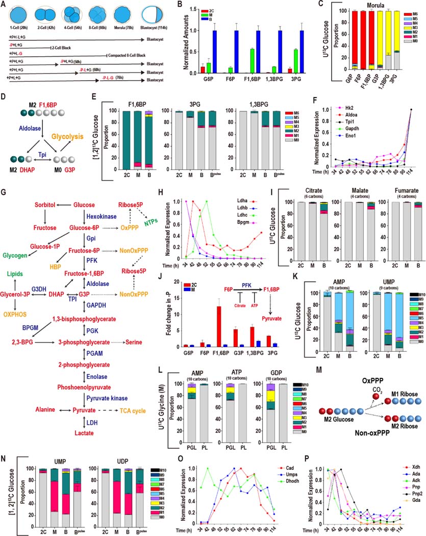
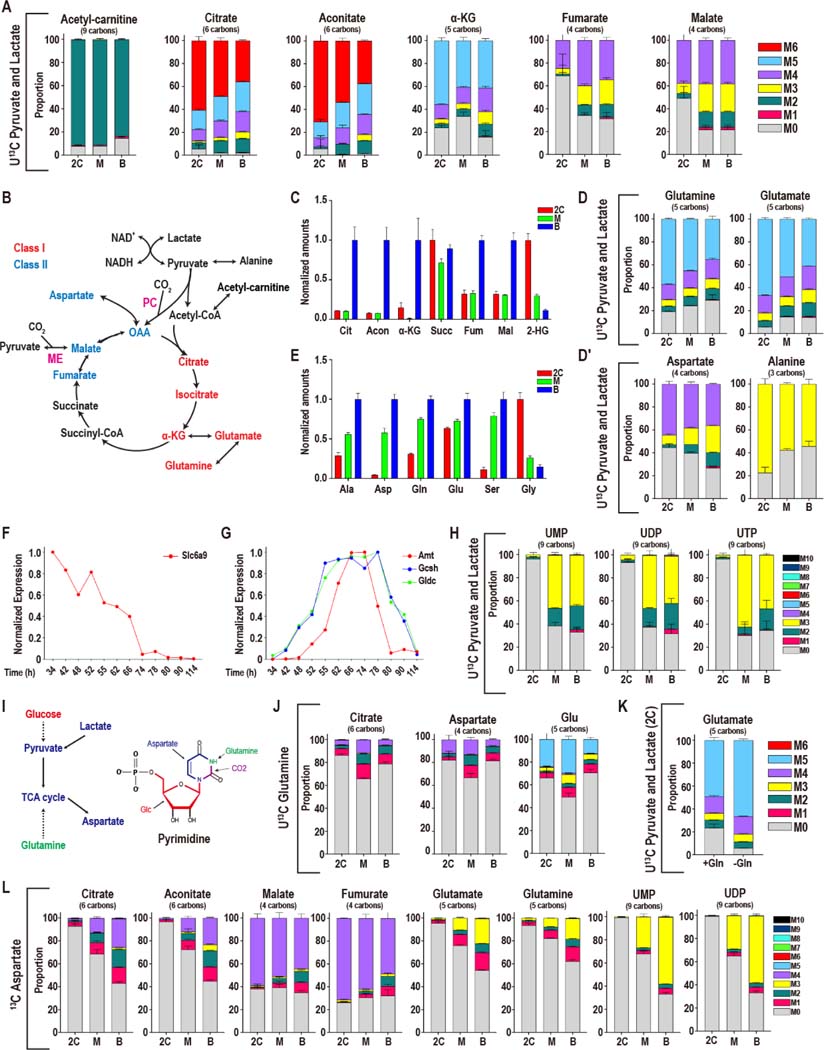
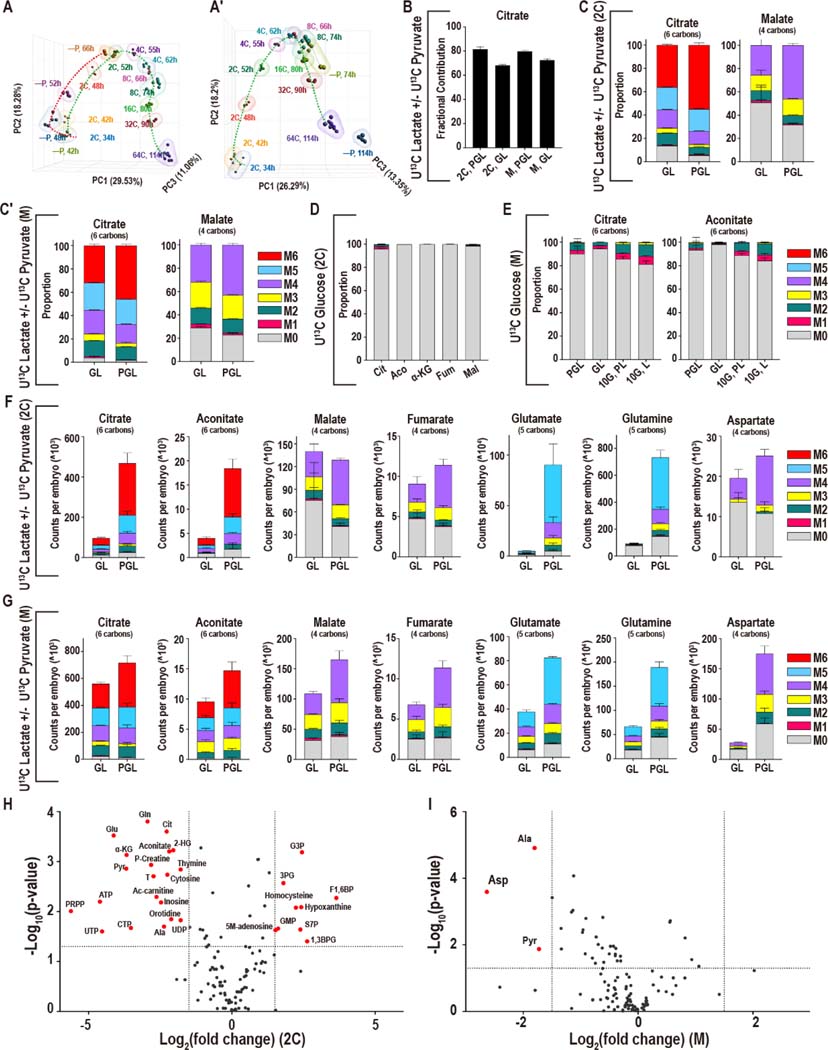
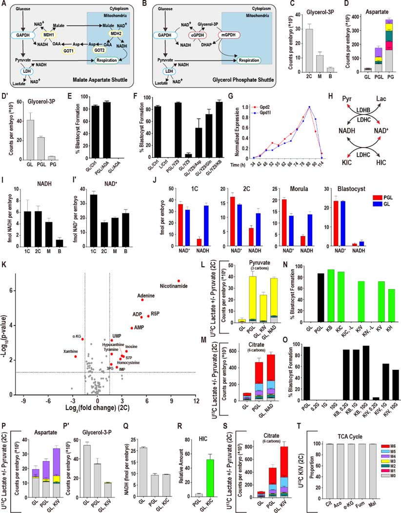
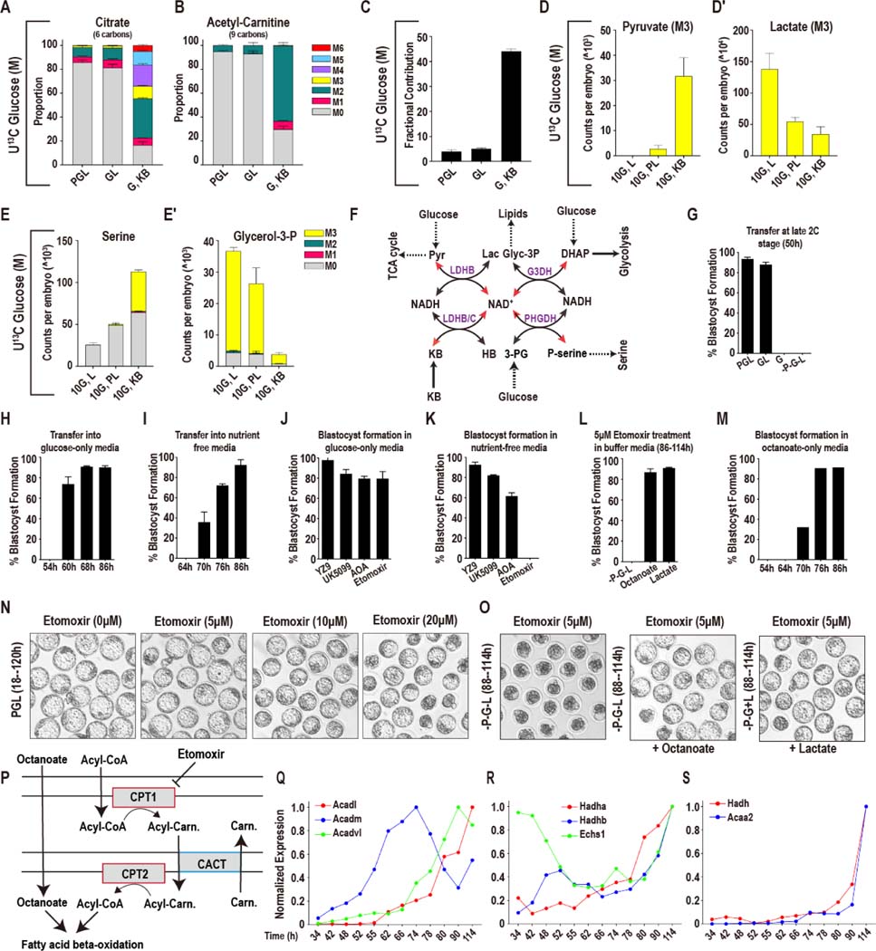
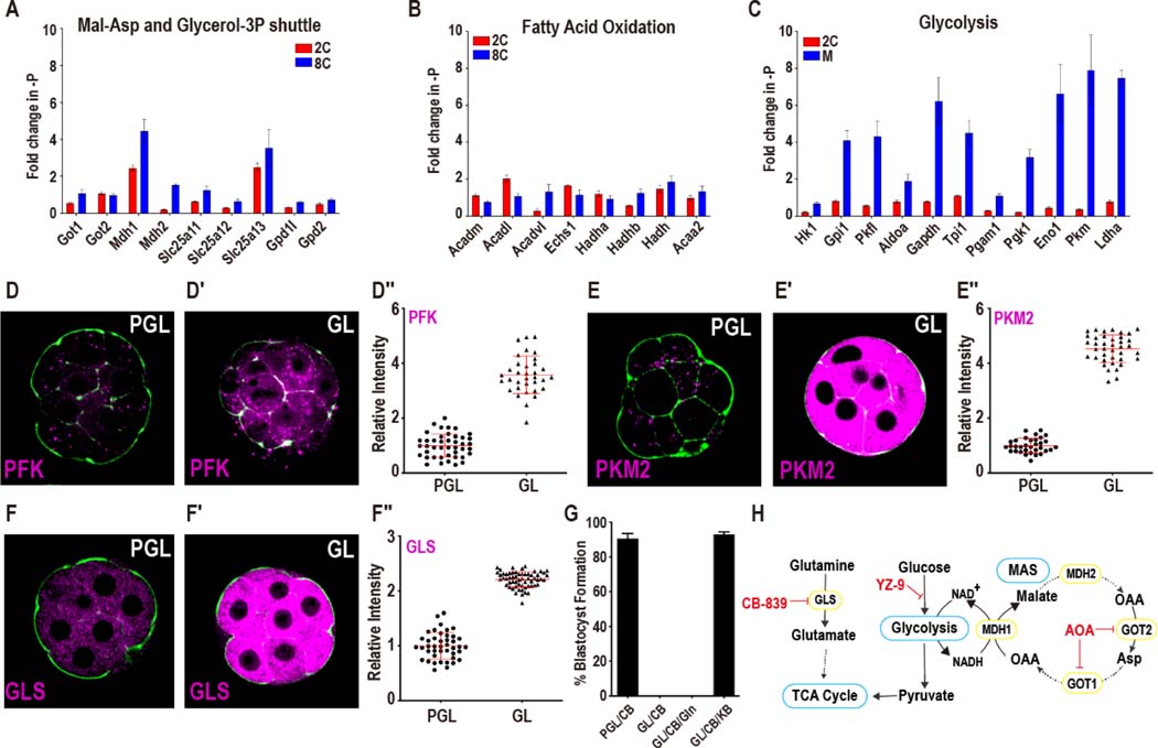
Mammalian preimplantation embryos follow a stereotypic pattern of development from zygotes to blastocysts. Here, we use labeled nutrient isotopologue analysis of small numbers of embryos to track downstream metabolites. Combined with transcriptomic analysis, we assess the capacity of the embryo to reprogram its metabolism through development. Early embryonic metabolism is rigid in its nutrient requirements, sensitive to reductive stress and has a marked disequilibrium between two halves of the TCA cycle. Later, loss of maternal LDHB and transcription of zygotic products favors increased activity of bioenergetic shuttles, fatty-acid oxidation and equilibration of the TCA cycle. As metabolic plasticity peaks, blastocysts can develop without external nutrients. Normal developmental metabolism of the early embryo is distinct from cancer metabolism. However, similarities emerge upon reductive stress. Increased metabolic plasticity with maturation is due to changes in redox control mechanisms and to transcriptional reprogramming of later-stage embryos during homeostasis or upon adaptation to environmental changes.

Keywords: MYC; NAD+/NADH; developmental metabolism; embryo; metabolic plasticity; metabolic reprogramming; preimplantation; redox; reductive stress; zygotic genome activation.


Conflict of interest statement

Declaration of interests The authors declare no competing interests.

Figures

Figure 1.
Figure 1.. Glucose metabolism during preimplantation development
In this and in all figures, times in hours (h) refer to time elapsed after human chorionic gonadotropin (hCG) injection that induces ovulation. Zygotes are isolated at 18–22h and cultured until the specified hours (h) post hCG. The metabolomic analysis is presented as scaled means ± SD and is obtained from 3 biological replicates. Each biological replicate contains more than 250 embryos. The RNA-seq analysis is presented as normalized values and is obtained from ~4 biological replicates of each of the 12 time-points that were analyzed. Each biological replicate contains RNA from 25 embryos. (A) A schematic representation of developmental progression of mouse preimplantation embryos in the presence of different combinations of nutrients. (B) Normalized amounts of glycolytic intermediates in 2C (48h), morula (M, 78h), and blastocysts (B, 96h). See Table S1 for non-normalized data. (C) U-13C glucose contributes carbon to the glycolytic intermediates at the morula stage. The lower glycolytic intermediates (1,3BPG and 3PG) have a prominent unlabeled component (gray). Gray: M0 unlabeled; colors: labeled as marked. Note that the “fully labeled” color depends on the total carbons in the metabolite (e.g., fully labeled is M6 (red) for G6P, M3 (yellow) for 1,3BPG and 3PG). (D) Schematic representing the fate of the 1 and 2 carbon of glucose at the aldolase reaction. (E) [1, 2]-13C glucose contributes carbon to the upper glycolytic intermediates (F1,6BP) at the 2C, morula (M) and blastocyst (B) stages. The lower glycolytic intermediates (1,3BPG and 3PG) are largely unlabeled, with an increased contribution at the blastocyst stage. For the blastocyst samples, labeled glucose is provided at the zygote stage (B) or 26 h prior to extraction (Bpulse). Colors: labeled as marked. (F, H) Expression of genes encoding glycolytic enzymes between early 2C (34h) and fully expanded blastocyst (114h) stages. (F) A majority of glycolytic enzymes increase in expression during later stages of development. (H) A subset is highest at the 2C stage and then declines. (G) Schematic of glucose metabolism. Red = metabolites, blue = enzymes, green = products, orange = pathways. (I) U-13C glucose only contributes minor amounts of carbon to TCA cycle metabolites, with a measurable contribution only at the blastocyst stage. (J) The lower glycolytic intermediates (G3P, 1,3BPG and 3PG) increase in abundance in 2C embryos following pyruvate withdrawal, but not in morula. Inset: Schematic illustration of the inhibition of PFK activity by citrate and ATP. (K) U-13C glucose contributes to nucleotide formation during development. The major isotopologue, M5 (blue) represents fully labeled ribose formation by glucose. (L) U-13C glycine contributes to purine base synthesis in morulae (M) if glucose (U-12C) is present (PGL), but does not do so when glucose is withheld (PL). (M) Schematic showing that when [1, 2]13C glucose is metabolized by the oxidative PPP, one labeled carbon is lost to CO2 formation and the ribose-5P formed is M1. When [1, 2]-13C glucose is metabolized by the non-oxidative PPP a carbon is not lost and the ribose-5P formed is M2. (N) [1, 2]13C glucose contributes carbon to the pyrimidine nucleotides UMP and UDP at the morula (M) and blastocyst (B) stages. M1 (pink) indicates ribose formation by the oxidative PPP, and M2 (green) ribose formation via the non-oxidative PPP. For the blastocyst samples, labeled glucose is provided at the zygote stage (B) or 26 h prior to extraction (Bpulse). (O, P) Expression of nucleotide metabolism genes. A majority of pyrimidine synthesis genes (O) increase in expression during the morula stage, whereas genes involved in purine degradation (P) peak in expression during the 2C stage. See also Figure S1 and Table S1.
Figure 2.
Figure 2.. Pyruvate and lactate metabolism during preimplantation development
(A) Isotopologue contributions of pyruvate and lactate to acetyl-carnitine and the TCA cycle metabolites. For acetyl-carnitine, M2 represents the acetyl group. Citrate and aconitate, but not fumarate and malate, are extensively labeled at all stages. The presence of M3 Class II metabolites suggests CO2 fixation via pyruvate carboxylase (PC) or malic enzyme (ME). In all figure panels, stages are represented as 2C: 2-cell, M: morula, B: blastocyst. (B) Schematic representing the TCA cycle and related amino acids. Class I metabolites are labeled red and Class II metabolites labeled blue. (C) Normalized amounts of TCA intermediates at each stage (see Table S1 for non-normalized amounts). (D, D′) Isotopologue contributions to the amino acids Glu, Gln, Ala and Asp. (E) Normalized amounts of non-essential amino acids at each stage (see Table S1 for non-normalized amounts). (F, G) The glycine transporter Slc6a9 (F) peaks in gene expression earlier than the enzymes of the glycine cleavage system (G). (H) U-13C pyruvate and U-13C lactate contribute to pyrimidine formation at later stages. (I) Schematic illustrating the role that glucose, pyruvate, lactate and glutamine play in forming aspartate and pyrimidine nucleotides. (J) U-13C Gln only contributes minor amounts of carbon to the TCA cycle and glutamate. (K) U-13C pyruvate and U-13C lactate generate a majority of Glu in 2C embryos both when exogenous unlabeled Gln (1 mM) is present (+Gln) or absent (-Gln). (L) In 2C embryos, U-13C Asp contributes to Class II metabolites, but not to Class I metabolites. In blastocysts, the Asp contribution to Class I metabolites increases. U-13C Asp is provided from the zygote stage and is present continuously until metabolites are extracted (see Figure S2J for “pulsed” labeling). See also Figure S2.
Figure 3.
Figure 3.. Adaptation to nutrient conditions
(A-A′) Principal component analysis (PCA) analysis of genes that are directly involved in metabolic processes (1947 genes) in embryos developing with and without pyruvate. Pyruvate is removed from the zygote stage (A) or the late 2C stage (A′). (B) In both 2C embryos and morulae, U-13C pyruvate/lactate contributes a majority of carbons to the total citrate pool in the presence (PGL) or absence (GL) of pyruvate. (C, C′) At both the 2C (C) and morula (C′) stages, the isotopologues of citrate that are formed from U-13C lactate alone (no pyruvate is present, GL) are similar to the isotopologues that are formed when both U-13C pyruvate and U-13C lactate (PGL) are present. (D, E) The contribution of U-13C glucose to the TCA cycle metabolites remains very low in 2C embryos (D) as well as in morulae (E) in GL media (lacking pyruvate), even when a high amount of glucose (10mM instead of the normal 0.2mM) is provided (10G, L). (F, G) Formation of Class I metabolites from U-13C lactate/pyruvate decreases when pyruvate is withheld from 2C embryos (F), but less so in morulae (G). Fumarate and malate are less sensitive, and Asp only decreases significantly in morulae. (H) In 2C embryos 20 metabolites decrease significantly, and 9 metabolites increase upon pyruvate withdrawal (GL). (I) In morulae, only Asp, pyruvate, and Ala decrease significantly. Metabolites are highlighted if P < 0.05 and the log2 fold-change > +/−1.5. See also Figure S3.
Figure 4.
Figure 4.. Mechanisms of metabolic plasticity
(A, B) Schematic of the Mal-Asp (A) and glycerol-3P shuttles (B). (C) The amount of glycerol-3P decreases between the 2C and blastocyst stages. (D) At the morula stage, Asp increases when lactate is absent (PG), and decreases when pyruvate is withheld (GL). (D′) Glycerol-3P decreases when lactate is absent and increases when pyruvate is withheld. (E) Following the 2C stage, inhibition of the Mal-Asp shuttle using 0.5 mM AOA does not impact the development of embryos cultured with pyruvate, lactate and glucose (PGL), but blocks development of embryos cultured in GL media. (F) The glycolysis inhibitor YZ-9 (2μM) blocks development of embryos deprived of pyruvate (GL) from the late 2C stage. This block is reversed by the addition of either Asp (1mM), Gln (1mM) or α-KB (1mM). (G) The glycerol-3P shuttle components Gpd1l and Gpd2 that are expressed in the embryo peak at the morula stage. (H) Schematic showing how pyruvate and lactate, and α-KIC and HIC, control NADH and NAD+ levels. (I-I′) NAD+ and NADH levels in 1C, 2C, morulae (M) and blastocysts (B). (I) NADH levels decrease during the development, whereas (I′) NAD+ falls markedly between the 1C and 2C stages then rises between the 2C and blastocyst stages. n = 4 biological replicates with ~100 embryos per replicate. (J) NAD+ and NADH levels in embryos grown with and without pyruvate at different stages of development. (K) In 2C embryos, provision of 5 mM NAD+ to pyruvate deprived embryos (GL, + 5 mM NAD+) restores the levels of many metabolites that fall when pyruvate is omitted from the media (compare with Figure 3H). A subset of metabolites increases including breakdown products of NAD+ (e.g., adenine, nicotinamide). Metabolites are highlighted if P < 0.05 and the log2 fold-change > +/−1.5. (L) 5 mM U-12C NAD+ or 1 mM U-12C KIV restores pyruvate levels in 2C embryos deprived of pyruvate by facilitating the use of U-13C lactate. (M) U-12C 5 mM NAD+ restores citrate levels in 2C embryos deprived of pyruvate by facilitating the use of U-13C lactate. The PGL (U-13C pyruvate and U-13C lactate) and GL (U-13C lactate) control samples are shared with Figure 3F, because these experiments were performed at the same time. (N) Alternative α-ketoacids (1mM), allow zygotes to develop into blastocysts in the absence of pyruvate, as long as lactate is provided. In a medium lacking pyruvate (GL), embryos do not form blastocysts unlike embryos that are cultured with pyruvate (PGL). The conditions represented by green bars all lack pyruvate (-P). The α-ketoacids KB, KIC, KIV, KV and KH all rescue (-P) embryos unless both pyruvate and lactate are absent (KIC, -L) (KIV, -L). Glucose (G) is present in all samples above, and pyruvate is absent in all samples except control (PGL). (O) Embryos that are transferred into pyruvate and lactate free media at the late 2C stage (after ZGA) (50h) are not viable over a range of glucose concentrations (0.2mM (0.2G), 1mM (1G) or 10mM (10G)) unless an α-ketoacid, such as KB or KIV, is provided. (P-P′) In 2C embryos, aspartate decreases and glycerol-3P increases in GL (U-13C lactate, U-12C glucose) media. KIV fully restores Asp levels and decreases the amount of glycerol-3P. The PGL (U-13C pyruvate and U-13C lactate) and GL (U-13C lactate) controls are shared with Figure 3F as these experiments were performed at the same time. (Q) NADH levels in zygotes with pyruvate, lactate and glucose (PGL), glucose and lactate (GL), or GL supplemented with 1mM KIC (GL, KIC). (R) Hydroxycaproate (HIC) increases in embryos that are provided with KIC. (S) U-12C KIV restores citrate levels in 2C embryos deprived of pyruvate by facilitating the use of U-13C lactate. The PGL (U-13C pyruvate and U-13C lactate) and GL (U-13C lactate) control samples are shared with Figure 3F, because these experiments were performed at the same time. (T) U-13C KIV does not contribute carbon to the TCA cycle in 2C embryos cultured in GL media. See also Figure S4.
Figure 5.
Figure 5.. Further metabolic plasticity in later stage embryos
(A, B) 10mM U-13C glucose contributes insignificant amounts of carbon to citrate (A) and acetyl-carnitine (B) in morulae in either PGL or GL media. In contrast, glucose contributes substantially with both pyruvate and lactate omitted if α-KB is provided. The control samples are shared with Figure 3E, because these experiments were performed at the same time. (C) For embryos cultured in PGL or GL, 10mM U-13C glucose contributes a minor amount of carbon to the total citrate pool. But with both pyruvate and lactate omitted, and α-KB provided, glucose contributes substantially to citrate. (D-E′) Metabolic reprogramming by α-ketoacid treatment. Pyruvate that is generated from glucose (M3) increases (D) in morula stage embryos when both pyruvate and lactate are omitted and α-KB is provided from the 2C stage. Lactate generated from glucose (M3) decreases (D′), serine synthesis from glucose increases (E) and glycerol-3P (E′) decreases. (F) Schematic illustrating how a change in the amount of pyruvate, lactate or α-KB that is provided to the embryo impacts NAD+ and NADH levels, and how this, in turn, impacts the activity of other metabolic pathways. (G) At the late 2C stage (50h) pyruvate-deprived embryos require both glucose and lactate to form blastocysts. (H) Glucose alone, without pyruvate and lactate, can support blastocyst formation after the mid-4C stage (60h). (I) After 70h (compacting morulae) embryos form blastocysts in the absence of all three nutrients (-P-G-L). (J) Embryos transferred from PGL into glucose-only media at 86h are insensitive to inhibition of glycolysis (YZ9, 2μM), pyruvate entry into the mitochondria (UK5099, 1μM), the Mal-Asp shuttle (AOA, 0.5mM) and fatty acid oxidation (FAO) (etomoxir, 5μM). (K) In the absence of all nutrients the embryos remain insensitive to inhibition of glycolysis, pyruvate transport, and the Mal-Asp shuttle, but are highly sensitive to FAO inhibition (etomoxir) and are unable to form blastocysts. (L, O) Both octanoate (200μM) and lactate (2.5mM) compensate for FAO inhibition (etomoxir, 5μM). (M) Pre-8C embryos do not form blastocysts in nutrient-free media supplemented with 200μM octanoate. (N) Inhibition of fatty acid oxidation (FAO; etomoxir, 5μM) does not inhibit blastocyst formation in normal PGL media. (P) Schematic illustrating the transport of octanoate (medium-chain) and long-chain fatty acids (called Acyl-CoA in Scheme) across the mitochondrial membranes, and representing how etomoxir inhibits long chain acyl-CoA transport, but not the transport of octanoate. (Q-S) Genes encoding enzymes of mitochondrial β-oxidation show high levels of expression in late morulae and blastocysts. See also Figure S5.
Figure 6.
Figure 6.. Metabolic response to reductive stress
(A, B) Genes in the Mal-Asp shuttle and glycerol-3P shuttle (A), and mitochondrial beta-oxidation (B) do not show a concerted increase when pyruvate is withdrawn. (C) The expression level of glycolytic genes increases in morulae, but not in 2C embryos. (D-E′′) Upon pyruvate withdrawal, protein levels of the glycolytic enzymes PFK (D-D′′) and PKM2 (E-E′′) show significant increases. Data is presented as the mean +/− standard deviation (n = ~25 embryos). (F-F′′) GLS protein increases when pyruvate is withheld. Data is presented as the mean +/− standard deviation (n = ~25 embryos). (G) Glutaminase inhibition (200nM CB-839) does not block the development of embryos in PGL media. Embryos in GL media, however, block in development following CB-839 treatment. α-KB (1mM) suppresses the block, but Gln (1mM) does not. (H) Schematic representing glycolysis, the Mal-Asp shuttle, glutamine metabolism and their inhibitors. See also Figure S6.
Figure 7.
Figure 7.. Mechanisms of metabolic reprogramming during reductive stress
(A-B′′) Myc protein localization upon pyruvate withdrawal in the morula and blastocyst. (A-A′′) In morulae, Myc is diffusely expressed in PGL (A), but is nuclearly localized in GL (A′). Quantitation in (A′′). (B-B′′) In blastocysts (114 h), Myc is expressed at low levels in PGL (B). Pyruvate withdrawal causes an increase in Myc expression in the ICM but not in the TE (B′). Quantitation in (B′′). Data is presented as the mean +/− standard deviation (n = ~25 embryos). (C-C′) Embryos cultured from the late 2C stage (50h) in GL (C′) but not in PGL (C) media block in development when treated with the Myc inhibitor KJ-Pyr-9 (abbreviated as KJ) at varying concentrations. (D) Embryos cultured in GL are sensitive to KJ (20nM). Gln (1mM) does not rescue, but supplementation with (1mM) α-KG (di-methyl) fully rescues the block. (E) Glycolytic genes do not increase in expression in pyruvate deprived embryos that are treated with 20nM KJ (green bars). The inhibitor-free sample (blue) is shared with Figure 6A, because these experiments were performed at the same time. (F-G′′′) Myc inhibition blocks the increase in pyruvate sensitive genes GLS (F-F′′′) and PKM2 (G-G′′′). GLS (F) and PKM2 (G) levels rise in GL media (F′, G′). KJ (20nM) prevents this increase in GLS (F′′) and in PKM2 (G′′). Quantitation in (F′′′, G′′′). Data is presented as the mean +/− standard deviation (n = ~25 embryos). (H-I′′) Myc functions under normal (PGL) growth conditions. Treatment with 20nM KJ (PGL medium, 2C, 50h) causes PKM2 (H, H′) and glutaminase (I, I′) protein levels to decrease. Quantitation in (H′′, I′′). Data is presented as the mean +/− standard deviation (n = ~25 embryos). (J-K′) A majority of Myc targets increase in expression at the morula stage (J) and more so in blastocysts (J′) following pyruvate withdrawal at the late 2C stage (50h). (K, K′) Myc targets do not increase in 2C embryos if they are cultured in a medium that lacks pyruvate from the zygote stage. See also Figure S7.

References

    1. Alexiou M, and Leese HJ (1992). Purine utilisation, de novo synthesis and degradation in mouse preimplantation embryos. Development 114, 185–192. - PubMed
    1. Alexiou M, and Leese HJ (1994). Enzymes of purine salvage and catabolism in the mouse preimplantation embryo measured by high performance liquid chromatography. J Reprod Fertil 101, 151–158. - PubMed
    1. Altman BJ, Stine ZE, and Dang CV (2016). From Krebs to clinic: glutamine metabolism to cancer therapy. Nat Rev Cancer 16, 749. - PubMed
    1. Aragon JJ, Tornheim K, Goodman MN, and Lowenstein JM (1981). Replenishment of citric acid cycle intermediates by the purine nucleotide cycle in rat skeletal muscle. Curr Top Cell Regul 18, 131–149. - PubMed
    1. Baltz JM (2001). Osmoregulation and cell volume regulation in the preimplantation embryo. Curr Top Dev Biol 52, 55–106. - PubMed

Publication types

LinkOut - more resources