. 2021 Aug 24;8(6):e1069.
doi: 10.1212/NXI.0000000000001069. Print 2021 Nov.

Melanoma Cell Adhesion Molecule Expressing Helper T Cells in CNS Inflammatory Demyelinating Diseases

Affiliations

Melanoma Cell Adhesion Molecule Expressing Helper T Cells in CNS Inflammatory Demyelinating Diseases

Ryotaro Ikeguchi et al. Neurol Neuroimmunol Neuroinflamm. .

Abstract

Background and objective: To elucidate the relationship between melanoma cell adhesion molecule (MCAM)-expressing lymphocytes and pathogenesis of CNS inflammatory demyelinating diseases (IDDs).

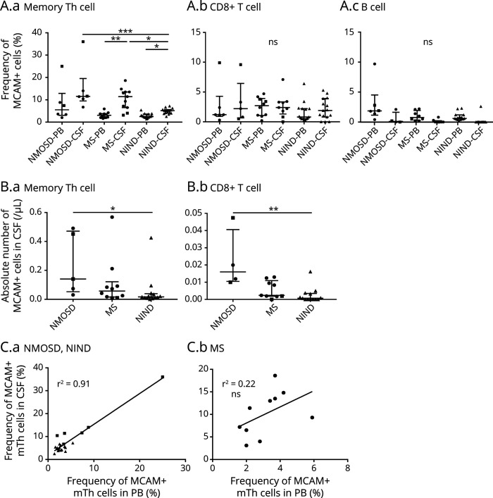
Methods: Patients with multiple sclerosis (MS) (n = 72) and neuromyelitis optica spectrum disorder (NMOSD, n = 29) were included. We analyzed the frequency and absolute numbers of MCAM+ lymphocytes (memory helper T [mTh] cells, naive helper T cells, CD8+ T cells, and B cells) in the peripheral blood (PB) and the CSF of patients with MS and NMOSD, treated with/without disease-modifying drugs (DMDs) or steroids, using flow cytometry.

Results: The frequency of MCAM+ cells was higher in the mTh cell subset than that in other lymphocyte subsets. A significant increase in the frequency and the absolute number of MCAM+ mTh cells was observed in the PB of patients with NMOSD, whereas no increase was observed in the PB of patients with MS. The frequency of CSF MCAM+ mTh cells was higher in relapsing patients with MS and NMOSD than that in the control group. Although there was no difference in the frequencies of MCAM+ lymphocytes among the DMD-treated groups, fingolimod decreased the absolute number of MCAM+ lymphocytes.

Discussion: MCAM+ mTh cells were elevated in the CSF of relapsing patients with MS and in both the PB and CSF of patients with NMOSD. These results indicate that MCAM contributes to the pathogenesis of MS and NMOSD through different mechanisms. MCAM could be a therapeutic target of CNS IDDs, and further study is needed to elucidate the underlying mechanism of MCAM in CNS IDD pathogenesis.


Figures

Figure 1
Figure 1. Frequency and Absolute Number of MCAM+ mTh Cells Increased in Patients With NMOSD
(A.a–A.c) Representative dot plot and histogram of MCAM+ (CD146+) mTh cells (CD3+ CD4+ CD45RA lymphocytes) using flow cytometry. The lymphocyte subsets included mTh cells, CD3+ CD4+ CD45RA lymphocytes; naive helper T cells, CD3+ CD4+ CD45RA+ lymphocytes; CD8+ T cells, CD3+ CD8+ lymphocytes; and B cells, CD3 CD19+ lymphocytes. The MCAM+ (CD146+) and α4-integrin+ (CD49d+) populations were defined based on the fluorescence intensity of the isotype control. (B) Frequency of MCAM+ cells in all lymphocyte subsets (n = 183: 72 patients with MS, 29 with NMOSD, 23 with MG, 34 with NIND, and 25 HCs). (C.a–C.d) Frequency of MCAM+ cells in all participants (72 patients with MS, 29 with NMOSD, 23 with MG, 34 with NIND, and 25 HCs). (D.a–D.c) Absolute number of MCAM+ cells in all patients (40 patients with MS, 23 with NMOSD, 17 with MG, and 26 NIND). Dimethyl fumarate (DMF)- (n = 11) and fingolimod- (n = 18) treated patients with MS were excluded because treatment with these DMDs reduces the absolute number of lymphocytes. Dots represent individual samples. The lower, upper, and middle lines correspond to the 25th centile, 75th centile, and median, respectively. *p < 0.05, **p < 0.01, and ***p < 0.001. HC = healthy control; MCAM = melanoma cell adhesion molecule; MG = myasthenia gravis; mTh cell = memory helper T cell; NIND = noninflammatory neurologic disorders; NMOSD = neuromyelitis optica spectrum disorder; ns = not significant.
Figure 2
Figure 2. Frequency and Absolute Number of MCAM+ Lymphocytes in DMD- or Steroid-Treated Patients With CNS IDD
(A.a–A.c) Frequency of MCAM+ cells in each lymphocyte subset among the DMD- or steroid-treated patients with MS (IFN-β [n = 12], DMF [n = 11], FTY [n = 18], and NTZ [n = 6]) and untreated patients with MS (n = 25). (B.a–B.c) Absolute number of MCAM+ cells in each lymphocyte subset among the DMD- or steroid-treated patients with MS (IFN-β [n = 9], DMF [n = 10], FTY [n = 18], and NTZ [n = 6]) and untreated patients (n = 24). (C) Frequency of MCAM+ mTh cells in the PB of patients with NMOSD and MG treated with or without PSL. At the time of examination, 25 of the 29 patients with NMOSD and 19 of the 23 patients with MG were undergoing treatment with oral PSL. Dots represent individual samples. The lower, upper, and middle lines correspond to the 25th centile, 75th centile, and median, respectively. *p < 0.05, **p < 0.01, and ***p < 0.001. DMD = disease-modifying drug; DMF = dimethyl fumarate; FTY = fingolimod; IDD = inflammatory demyelinating disease; IFN = interferon; MCAM = melanoma cell adhesion molecule; MG = myasthenia gravis; mTh cell = memory helper T cell; NMOSD = neuromyelitis optica spectrum disorder; ns = not significant; NTZ = natalizumab; PB = peripheral blood; PSL = prednisolone.
Figure 3
Figure 3. Frequency of MCAM+ mTh Cells Was Increased in the CSF of Patients With CNS IDD
(A.a–A.c) Frequency of CSF and PB MCAM+ lymphocytes (MS [n = 10], NMOSD [n = 6], and NIND [n = 15]). At examination, 8 patients with MS and 4 patients with NMOSD were in the relapse phase. (B.a–B.b) Absolute number of CSF MCAM+ lymphocytes (MS [n = 10], NMOSD [n = 5], and NIND [n = 13]). The absolute number of MCAM+ B cells is not shown in this figure because this subset has been rarely observed in the CSF. Closed circles represent the relapse phase, and closed squares represent the remission phase in (A) and (B). (C) Correlation between the frequencies of PB and CSF MCAM+ mTh cells in patients with (C.a) NMOSD (n = 6, closed square) and NIND (n = 15, closed triangle) and (C.b) only in patients with MS (n = 10, closed circle). Dots represent individual samples. The lower, upper, and middle lines correspond to the 25th centile, 75th centile, and median, respectively. *p < 0.05, **p < 0.01, and ***p < 0.001. IDD = inflammatory demyelinating disease; MCAM = melanoma cell adhesion molecule; mTh cell = memory helper T cell; NIND = noninflammatory neurologic disorders; NMOSD = neuromyelitis optica spectrum disorder; ns = not significant; PB = peripheral blood.

References

    1. McFarland HF, Martin R. Multiple sclerosis: a complicated picture of autoimmunity. Nat Immunol. 2007;8(9):913-919. - PubMed
    1. Dendrou CA, Fugger L, Friese MA. Immunopathology of multiple sclerosis. Nat Rev Immunol. 2015;15(9):545-558. - PubMed
    1. Panitch HS, Hirsch RL, Schindler J, Johnson KP. Treatment of multiple sclerosis with gamma interferon: exacerbations associated with activation of the immune system. Neurology. 1987;37(7):1097-1102. - PubMed
    1. Tzartos JS, Friese MA, Craner MJ, et al. . Interleukin-17 production in central nervous system-infiltrating T cells and glial cells is associated with active disease in multiple sclerosis. Am J Pathol. 2008;172(1):146-155. - PMC - PubMed
    1. Kebir H, Ifergan I, Alvarez JI, et al. . Preferential recruitment of interferon-gamma-expressing TH17 cells in multiple sclerosis. Ann Neurol. 2009;66(3):390-402. - PubMed

Publication types

MeSH terms