. 2021 Aug 24;11(1):17094.
doi: 10.1038/s41598-021-96524-z.

Kinetic studies and homology modeling of a dual-substrate linalool/nerolidol synthase from Plectranthus amboinicus

Affiliations

Kinetic studies and homology modeling of a dual-substrate linalool/nerolidol synthase from Plectranthus amboinicus

Nur Suhanawati Ashaari et al. Sci Rep. .

Abstract

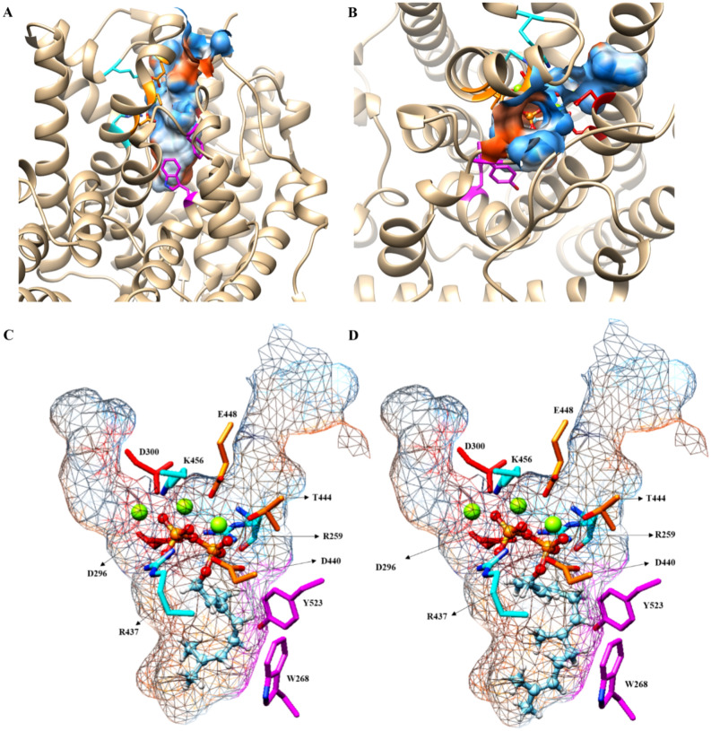
Linalool and nerolidol are terpene alcohols that occur naturally in many aromatic plants and are commonly used in food and cosmetic industries as flavors and fragrances. In plants, linalool and nerolidol are biosynthesized as a result of respective linalool synthase and nerolidol synthase, or a single linalool/nerolidol synthase. In our previous work, we have isolated a linalool/nerolidol synthase (designated as PamTps1) from a local herbal plant, Plectranthus amboinicus, and successfully demonstrated the production of linalool and nerolidol in an Escherichia coli system. In this work, the biochemical properties of PamTps1 were analyzed, and its 3D homology model with the docking positions of its substrates, geranyl pyrophosphate (C10) and farnesyl pyrophosphate (C15) in the active site were constructed. PamTps1 exhibited the highest enzymatic activity at an optimal pH and temperature of 6.5 and 30 °C, respectively, and in the presence of 20 mM magnesium as a cofactor. The Michaelis-Menten constant (Km) and catalytic efficiency (kcat/Km) values of 16.72 ± 1.32 µM and 9.57 × 10⁻³ µM-1 s-1, respectively, showed that PamTps1 had a higher binding affinity and specificity for GPP instead of FPP as expected for a monoterpene synthase. The PamTps1 exhibits feature of a class I terpene synthase fold that made up of α-helices architecture with N-terminal domain and catalytic C-terminal domain. Nine aromatic residues (W268, Y272, Y299, F371, Y378, Y379, F447, Y517 and Y523) outlined the hydrophobic walls of the active site cavity, whilst residues from the RRx8W motif, RxR motif, H-α1 and J-K loops formed the active site lid that shielded the highly reactive carbocationic intermediates from the solvents. The dual substrates use by PamTps1 was hypothesized to be possible due to the architecture and residues lining the catalytic site that can accommodate larger substrate (FPP) as demonstrated by the protein modelling and docking analysis. This model serves as a first glimpse into the structural insights of the PamTps1 catalytic active site as a multi-substrate linalool/nerolidol synthase.


Conflict of interest statement

The authors declare no competing interests.

Figures

Figure 1
Figure 1
Biochemical characterization of PamTps1. (A) pH; (B) Temperature; (C) Mg2+ concentrations; (D) Mn2+ concentrations. Michaelis–Menten plot of PamTps1 at different concentrations of (E) Mn2+; (F) Mg2+; (G) GPP and (H) FPP. The saturation curve was constructed using Michaelis–Menten equation by hyperbolic regression. Values were reported as the mean of relative activity ± SD of triplicate analysis.
Figure 2
Figure 2
Protein homology modelling of PamTps1 using SWISS-MODEL server showed the structural domains and the active site of the enzyme. (A) model structure of PamTps1 made up of α-helices with N-terminal domain (green) and C-terminal domain (blue). (B) Ribbon view of PamTps1 model. The helical segment was designated according to Tarshis et al.. All conserved motifs were labelled in the figure and Mg2+ was illustrated as green spheres.
Figure 3
Figure 3
Superimposition of PamTps1 model (purple) with BPPS template (brown) using Chimera. The α-carbon RMSD value of 0.203 Å indicated the two structures were exceptionally similar. The aspartate-rich motif was red, the DTE motif was orange and the green spheres were magnesium ions.
Figure 4
Figure 4
Ligand-PamTps1 interactions. Three-dimensional (3D) view of (A) GPP and (B) FPP docking at PamTps1 active site. The red–orange balls and stick chains represent PPi, green spheres are the Mg2+, the red side chains are the DDxxD motif, the orange side chains are the DTE motifs, the blue side chains are the hydrogen bond donor residues, the magenta side chains represent the aromatic residues and the cyan lines are the hydrogen bonds. Two-dimensional (2D) view of (C) GPP and (D) FPP docking at PamTps1 active site. Hydrogen bonds are shown as green dotted lines, Mg2+ are shown in green spheres and spoked arcs represent residues making non-bonded contacts with the hydrophobic tail of the ligand. Details of the docking result are summarized in Table S2.
Figure 5
Figure 5
The PamTps1 active site pocket. The overview of PamTps1 active site pocket from (A) top and (B) side views. Docking positions of (C) GPP and (D) FPP in PamTps1 active site cavity. The PamTps1 active site is a deep hydrophobic pocket consisting of C, D, F, G, H and J helices. The red–orange ball and stick chains represent the PPi of substrate, green spheres are the Mg2+, the orange side chains are the DTE motifs, the blue side chains are the hydrogen bond donor residues and the magenta side chains represent the aromatic residues.

References

    1. Tholl, D. Biosynthesis and biological functions of terpenoids in plants in Biotechnology of isoprenoids (eds. Schrader, J. & Bohlmann, J.) 63-106 (Springer, 2015). - PubMed
    1. George, K. W., Alonso-Gutierrez, J., Keasling, J. D. & Lee, T. S. Isoprenoid drugs, biofuels, and chemicals—artemisinin, farnesene, and beyond in Biotechnology of isoprenoids (eds. Schrader, J. & Bohlmann, J.) 355–389 (Springer, 2015). - PubMed
    1. Turner G, Gershenzon J, Nielson EE, Froehlich JE, Croteau R. Limonene synthase, the enzyme responsible for monoterpene biosynthesis in peppermint, is localized to leucoplasts of oil gland secretory cells. Plant Physiol. 1999;120:879–886. doi: 10.1104/pp.120.3.879. - DOI - PMC - PubMed
    1. Bohlmann J, Meyer-Gauen G, Croteau R. Plant terpenoid synthases: Molecular biology and phylogenetic analysis. Proc. Natl. Acad. Sci. 1998;95:4126–4133. doi: 10.1073/pnas.95.8.4126. - DOI - PMC - PubMed
    1. Whittington DA, et al. Bornyl diphosphate synthase: Structure and strategy for carbocation manipulation by a terpenoid cyclase. Proc. Natl. Acad. Sci. 2002;99:15375–15380. doi: 10.1073/pnas.232591099. - DOI - PMC - PubMed

Publication types

MeSH terms