. 2021 Aug 14:16:5495-5512.
doi: 10.2147/IJN.S318331. eCollection 2021.

Identification of Novel Ligands for Targeted Antifibrotic Therapy of Chronic Pancreatitis

Affiliations

Identification of Novel Ligands for Targeted Antifibrotic Therapy of Chronic Pancreatitis

Jessica Hung et al. Int J Nanomedicine. .

Abstract

Purpose: Chronic pancreatitis (CP) is an inflammatory disorder of the pancreas that leads to impaired pancreatic function. The limited therapeutic options and the lack of molecular targeting ligands or non-serum-based biomarkers hinder the development of target-specific drugs. Thus, there is a need for an unbiased, comprehensive discovery and evaluation of pancreatitis-specific ligands.

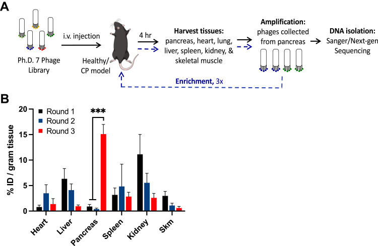
Methods: This study utilized a computational-guided in vivo phage display approach to select peptide ligands selective for cellular components in the caerulein-induced mouse model of CP. The identified peptides were conjugated to pegylated DOPC liposomes via the reverse-phase evaporation method, and the in vivo specificity and pharmacokinetics were determined. As proof of concept, CP-targeted liposomes were used to deliver an antifibrotic small molecular drug, apigenin. Antifibrotic effects determined by pancreatic histology, fibronectin expression, and collagen deposition were evaluated.

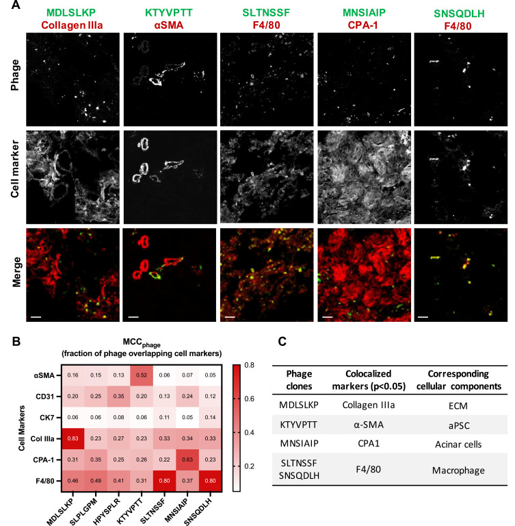
Results: We have identified five peptides specific for chronic pancreatitis and demonstrated selectivity to activated pancreatic stellate cells, acinar cells, macrophages, and extracellular matrix, respectively. MDLSLKP-conjugated liposomes demonstrated an increased particle accumulation by 1.3-fold in the inflamed pancreas compared to the control liposomes. We also observed that targeted delivery of apigenin resulted in improved acini preservation, a 37.2% and 33.1% respective reduction in collagen and fibronectin expression compared to mice receiving the free drug, and reduced oxidative stress in the liver.

Conclusion: In summary, we have developed a systematic approach to profile peptide ligands selective for cellular components of complex disease models and demonstrated the biomedical applications of the identified peptides to improve tissue remodeling in the inflamed pancreas.

Keywords: drug delivery; next-generation sequencing; peptide ligands; phage display; targeted liposomes.


Conflict of interest statement

KAK is the CEO and Founder of ZielBio, Inc. ZielBio played no role in funding this work. KAK and JH report a patent HIGH-THROUGHPUT ANALYTIC SELECTIONS OF NOVEL TARGETING LIGANDS FOR CHRONIC PANCREATITIS AND DEVELOPMENT OF TARGET-SPECIFIC LIPOSOMES FOR ANTIFIBROTIC THERAPY pending to United States Patent and Trademark Office. The application was filed on 2/5/2021. ALK is a subcontract of an NIH grant to SoundPipe Therapeutics, a company in the area of microbubble-assisted drug delivery. The authors report no other conflicts of interest in this work.

Figures

Figure 1
Figure 1
Pharmacokinetics of peptide-modified liposomes. (A) Table showing characteristic features of surface-modified liposomes with peptides identified to target collagen IIIa+ and acinar cells. (B) In vivo IVIS images of CP mice over 72h time course post-injection of peptide-modified liposomes. The white arrow indicates fluorescent signals detected at the pancreas region. (C) Ex vivo IVIS images of the pancreas at 4, 48, and 72h post liposome injection. (D) Biodistribution of DiD-labeled liposomes in CP mice at 48h post injection. Fluorescent intensity is normalized to the number of particles injected, the number of DiD per liposome, and the mass of the pancreas. N = 3. Student’s t-test was used to compare the targeted liposomes to the no peptide liposomes. *p < 0.05.
Figure 2
Figure 2
In vivo phage screening in the chronic pancreatitis mouse model. (A) A schematic of the in vivo phage biopanning process to screen for clones specific for CP pancreas. (B) Phage titering of in vivo phage screening in caerulein-induced CP mice. Phage pools (% injected dose per gram tissue) were recovered from the pancreas and various organs in 3 rounds of the biopanning process. One-way ANOVA and Tukey–Kramer tests were used to compare round 3 vs round 1, and round 3 vs round 2. N = 3; ***p<0.0001.
Figure 3
Figure 3
In silico selection and in vivo validation of CP targeting clones. CP targeting candidate clones were selected based on (A) PHASTpep, (B) replicability between 3 mice (asterisk indicates clones that have not been identified as ligands for known targets), (C) clone enrichment between 3 rounds of biopanning, and (D) motif clustering analysis. The normalized frequency count of each clone is represented in heatmaps. (E) Phage clone specificity validation by homology groups revealed preferential bindings of Group 2 and 3 targeting clones to CP pancreas over the healthy pancreas. N = 5. Mean ± SEM. A Student’s t-test was used to compare the targeting phage (VT680)-to-wild type phage (VT750) ratio in CP versus the same ratio in the healthy pancreas. *p-value < 0.05 (p = 0.0275 for Group 2; p = 0.0443 for Group 3). (F) Phage clones from Group 2 and 3 were validated individually in both healthy and CP mice. Fold change represents the ratio of targeting-to-wild type ratio in CP over healthy mice. 7/9 clones showed higher phage accumulation in CP over healthy pancreas (fold change >1).
Figure 4
Figure 4
CP-homing phage clones show selectivity for cellular components in the CP pancreas. (A) Immunofluorescence images of VT680-labeled phage colocalized with cell markers in the inflamed pancreas. Six cell markers were stained to represent six common cellular components in CP: αSMA (activated PSC), CD31 (endothelium), CK7 (epithelium), Collagen IIIa (ECM), CPA-1 (acinar cells), and F4/80 (macrophages). Colors in the merged images represent phage (green) and cell markers (red). Scale bar: 20 μm. (B) Heatmap of mean Manders’ correlation coefficient (MCC) representing the fraction of phage overlapping with the cell markers. Manders’ colocalization analysis was performed using the ImageJ plug-in JACoP. N = 10–12 images per marker, per clone. Col IIIa: collagen IIIa. (C) A table summarizing phage clones shown statistically significant selectivity for a single cellular component in CP. One-way ANOVA and Tukey–Kramer tests were used to compare the MCC of all cell markers for each clone. The result was considered significant if the p-value ≤ 0.05.
Figure 5
Figure 5
Immunofluorescence of peptide-conjugated liposomes in CP pancreas. (A) NC liposomes were non-specifically taken up by macrophages present in the inflamed pancreas. MDLSLKP liposomes colocalized with extracellular matrix (collagen IIIa+ cells). Color code: green for liposome (DiD) and red for cell markers. Scale bar: 20 μm. (B) Box-and-whisker plot of MCC values of liposomes overlapping cell markers. Liposome selectivity for the corresponding cell types was analyzed using the ImageJ plug-in JACoP. N = 7~12 images per group. One-way ANOVA and Tukey–Kramer tests were used to compare the MCC of all cell markers for each liposome. *p < 0.05. No statistically significant difference was observed in spatial localization of MNSIAIP liposomes with any stained cell types.
Figure 6
Figure 6
Targeted delivery of Apigenin reduces fibrosis. (A) Schematic of CP mouse model followed by 3-week treatments of either empty ECM liposome (vehicle), free Apigenin (free drug), Apigenin-encapsulated naked liposomes (Api-Naked Lip), or Apigenin-encapsulated MDLSLKP liposomes (Api-ECM Lip) (N=5). (B) H&E staining of pancreas by the end of 3-week treatments. Reduced interstitial space and acinar atrophy (indicated by arrows) were observed in the pancreas treated by targeted liposomes compared to free drug and the control liposomes. Scale bar: 50 μm. (C) Pancreas immuno-stained for fibronectin demonstrated targeted delivery of Apigenin significantly decreased fibronectin expression. Scale bar: 100 μm. (D) Picrosirius red staining of the pancreas. Scale bar: 50 μm. (E) Number of acini atrophy found in a 256 μm x 256 μm image. N = 5 images/animal, 3 animals/group. (F) Quantification of fibronectin-positive area. N = 8 images/animal, 5 animals/group. (G) Quantification of Picrosirius red-positive area. N = 12 images/animal, 5 animals/group. In all images, ANOVA and Tukey’s test were used to compare Api-ECM Lip to the rest of the treatment groups. *p<0.05, ****p<0.0001.

Similar articles

Cited by

References

    1. Gardner TB, Adler DG, Forsmark CE, Sauer BG, Taylor JR, Whitcomb DC. ACG clinical guideline: chronic pancreatitis. Off J Am Coll Gastroenterol ACG. 2020;115(3):322–339. doi:10.14309/ajg.0000000000000535 - DOI - PubMed
    1. Beyer G, Habtezion A, Werner J, Lerch MM, Mayerle J. Chronic pancreatitis. Lancet Lond Engl. 2020;396(10249):499–512. doi:10.1016/S0140-6736(20)31318-0 - DOI - PubMed
    1. Morgan K, Owczarski SM, Borckardt J, et al. Quality of life after pancreatectomy with islet autotransplantation for chronic pancreatitis. J Gastrointest Surg. 2012;16(1):129–134. doi:10.1007/s11605-011-1744-y - DOI - PubMed
    1. Machicado JD, Amann ST, Anderson MA, et al. Quality of life in chronic pancreatitis is determined by constant pain, disability/ unemployment, current smoking, and associated co-morbidities. Am J Gastroenterol. 2017;112(4):633–642. doi:10.1038/ajg.2017.42 - DOI - PMC - PubMed
    1. Han S, Patel B, Min M, et al. Quality of life comparison between smokers and non-smokers with chronic pancreatitis. Pancreatol off J Int Assoc Pancreatol IAP Al. 2018;18(3):269–274. doi:10.1016/j.pan.2018.02.012 - DOI - PMC - PubMed