

Breast metastases from primary lung cancer: a retrospective case series on clinical, ultrasonographic, and immunohistochemical features
- PMID: 34430360
- PMCID: PMC8350075
- DOI: 10.21037/tlcr-21-542
Breast metastases from primary lung cancer: a retrospective case series on clinical, ultrasonographic, and immunohistochemical features
Abstract
Background: Lung cancer metastases to the breast are less common and consequently have received much less attention in clinical practice. The purpose of this study was to provide a better understanding of clinical, ultrasonographic, and immunohistochemical features of breast metastases from primary lung cancer.
Methods: This retrospective case series included patients with breast metastases from primary lung cancer between January 2012 and December 2020. Clinical features, ultrasonographic characteristics, and immunohistochemical findings were evaluated in this analysis.
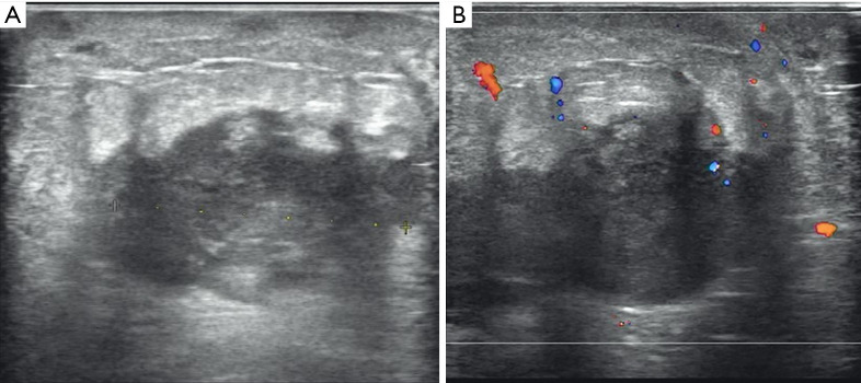
Results: In all, 7 cases (mean ± standard deviation age: 57.4±8.3 years; range, 49-70 years) were evaluated. The maximum size of breast lesions in 6 cases ranged from 1.2 to 4.5 cm, while 1 case showed a diffused pattern. Ultrasound features of breast metastases from lung cancer were irregular (5/7, 71.4%), indistinct (6/7, 85.7%), hypoechoic (7/7, 100.0%), and parallel (6/7, 85.7%) masses without calcification. Immunohistochemical staining test was positive for thyroid transcription factor 1 (TTF-1) in all patients (7/7, 100.0%), 3 cases (3/5, 60.0%) were negative for p63, 5 cases (5/5, 100.0%) were positive for cytokeratin 7 (CK7), 4 cases (4/5, 80.0%) were positive for napsin A.
Conclusions: The ultrasonographic features of lung metastases to the breast are clinically important to understand. A known history of the primary lung cancer is of great importance when evaluating patients with a breast nodule. The presence of an ipsilateral lung cancer, breast nodule and axillary lymphadenopathy should be considered with pathological and immunohistochemical data to differentiate breast metastases from a primary breast malignancy in this setting.
Keywords: Breast metastases; case series; immunohistochemistry; lung cancer; ultrasonography.
2021 Translational Lung Cancer Research. All rights reserved.
Conflict of interest statement
Conflicts of Interest: All authors have completed the ICMJE uniform disclosure form (available at https://dx.doi.org/10.21037/tlcr-21-542). The authors have no conflicts of interest to declare.
Figures


References
Grants and funding
LinkOut - more resources
Full Text Sources
Research Materials