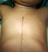
Successful surgical repair of a sternum cleft using composite mesh: A case report and new technical note
- PMID: 34430867
- PMCID: PMC8327679
- DOI: 10.7196/AJTCCM.2021.v27i2.103
Successful surgical repair of a sternum cleft using composite mesh: A case report and new technical note
Abstract
Sternal clefts are infrequent congenital malformations, particularly in their complete presentation. There are less than 100 descriptions of these defects published in the literature worldwide. We report a clinical case of lower sternal cleft associated with congenital laparoschisis in a 2-year-old boy. Surgery was performed because of recurrent pneumopathy and the risk of cardiorespiratory decompensation in the midterm. A semi-resorbable prosthesis was used for sternal closure. We have not observed any complications with this sternal closure system in our patient. This approach is easy, safe, effective and not harmful to a child's growth.
Keywords: bifid sternum; chest wall deformity; congenital abnormality; laparoschisis; pentalogy of Cantrell syndrome; sternal clef.
Conflict of interest statement
Conflicts of interest: None.
Figures


References
-
- Demircan M, Gürbüz N, Karaman A. A bifid sternum case underwent the earliest repair in the literature. İnönü Üniversitesi Tıp Fakültesi Dergisi. 2010;12(4):253–255.
-
- Chellaoui M, Chat L, Achaabal F, et al. Fissure sternale congénitale totale à propos d’un cas. Médecine du Maghreb. 2001;90(3):16–18.
Publication types
LinkOut - more resources
Full Text Sources
