. 2021 Aug 13;2(3):100743.
doi: 10.1016/j.xpro.2021.100743. eCollection 2021 Sep 17.

Electrophysiological and anatomical characterization of synaptic remodeling in the mouse whisker thalamus

Affiliations

Electrophysiological and anatomical characterization of synaptic remodeling in the mouse whisker thalamus

Yoshifumi Ueta et al. STAR Protoc. .

Abstract

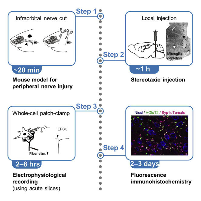
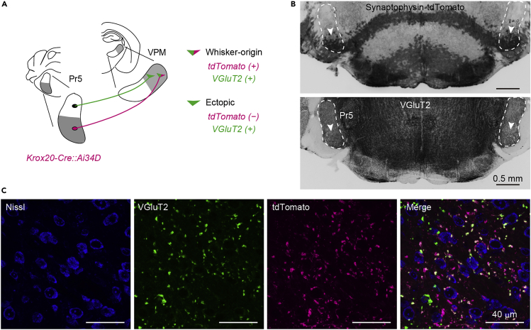
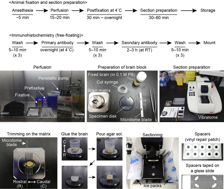
In the central nervous system, developmental and pathophysiologic conditions cause a large-scale reorganization of functional connectivity of neural circuits. Here, by using a mouse model for peripheral sensory nerve injury, we present a protocol for combined electrophysiological and anatomical techniques to identify neural basis of synaptic remodeling in the mouse whisker thalamus. Our protocol provides comprehensive approaches to analyze both structural and functional components of synaptic remodeling. For complete details on the use and execution of this protocol, please refer to Ueta and Miyata, (2021).

Keywords: cell biology; microscopy; model organisms; neuroscience.


Conflict of interest statement

The authors declare no competing interests.

Figures

None
Graphical abstract
Figure 1
Figure 1
Development of mouse model for the infraorbital nerve (ION) cut (A) A setup for mouse surgery. (B) Exposed ION. Scale bar, 5 mm. (C) No running ION after ION cut (IONC). Scale bar, 5 mm.
Figure 3
Figure 3
Focal injection using iontophoresis (A) A retrograde tracer, Alexa Fluor 647-conjugated cholera toxin subunit B (CTB647, 0.2% in ddH2O), is iontophoretically delivered to the dorsal part of the thalamic ventral posteromedial nucleus (VPM). VPM is identified by VGLUT2 immunohistochemistry using a guinea pig anti-VGluT2 antibody and an Alexa Fluor 488-conjugated secondary antibody. Gray-scale images are acquired using an epifluorescence microscope (Axio Scope.A1, Carl Zeiss) equipped with a cooled CCD camera (QSI RS 6.1, Quantum Scientific Imaging) and pseudocolored using Adobe Photoshop software. D, dorsal; L, lateral; VPL, ventral posterolateral nucleus. Scale bar, 0.5 mm. (B) Retrogradely labeled cells in the ventral part of the principal trigeminal nucleus of the brainstem (Pr5) in the contralateral hemisphere. M, medial. Scale bar, 0.5 mm.
Figure 2
Figure 2
The timing and workflow of stereotaxic injection
Figure 4
Figure 4
Local and brain-wide depletion of microglia (A) Clodronate liposomes, which induce apoptosis of microglia, are locally injected to the brainstem using a Hamilton syringe. No immunoreactivity against a myeloid cell marker Iba1 is found in the injection site. Immunohistochemistry is performed using a rabbit anti-Iba1 antibody and an Alexa Fluor 488-conjugated secondary antibody. Gray-scale images are acquired using the epifluorescence microscope same as in Figure 3 and inverted using Adobe Photoshop software. Scale bar, 0.5 mm (left) or 0.2 mm (right). (B) Brain-wide microglia are depleted by systemic administration of PLX3397, which inhibits microglial proliferation. An arrow indicates a remaining Iba1-positive cell in the brainstem. Scale bar, 0.2 mm.
Figure 6
Figure 6
A setup for whole-cell patch clamp recording and example experiments (A) A setup for patch-clamp experiments. (B) Estimation of innervated axon numbers by excitatory postsynaptic current (EPSC) recording from a thalamic VPM neuron under voltage-clamp.
Figure 5
Figure 5
The timing and workflow of preparation of acute brain slices
Figure 7
Figure 7
Post hoc visualization of cell morphology after electrophysiological recording A VPM neuron is visualized with Alexa Fluor 488-conjugated streptavidin. Immunohistochemistry against vesicular glutamate transporter type 2 (VGluT2) and Alexa Fluor 647-conjugated secondary antibody is simultaneously performed. Gray-scale images are acquired using a fluorescent microscope (BZ-X810, Keyence) and pseudocolored. The slice is resectioned at 40 μm. Scale bar, 20 μm.
Figure 8
Figure 8
Schematic illustrating the identification of pathway-specific axon terminals in the thalamus and example sections (A) Identification of whisker-origin and ectopic (non-whisker-origin) axon terminals in the thalamic VPM using Krox20-Cre mice crossed with Ai34D mice, which express synaptophysin-tdTomato under Cre. (B) Cre recombinase is expressed in principal neurons in the ventral whisker region of Pr5. Scale bar, 0.5 mm. (C) Axon terminals in VPM. Gray-scale images are acquired using a confocal laser scanning microscope (LSM710, Carl Zeiss) and pseudocolored. Scale bar, 40 μm.
Figure 9
Figure 9
The timing and workflow of immunohistochemistry using fixed brain sections

References

    1. Bechara A., Laumonnerie C., Vilain N., Kratochwil C.F., Cankovic V., Maiorano N.A., Kirschmann M.A., Ducret S., Rijli F.M. Hoxa2 selects barrelette neuron identity and connectivity in the mouse somatosensory brainstem. Cell Rep. 2015;13:783–797. - PubMed
    1. Eguchi K., Velicky P., Hollergschwandtner E., Itakura M., Fukazawa Y., Danzl J.G., Shigemoto R. Advantages of acute brain slices prepared at physiological temperature in the characterization of synaptic functions. Front. Cell. Neurosci. 2020;14:63. - PMC - PubMed
    1. Elmore M.R., Najafi A.R., Koike M.A., Dagher N.N., Spangenberg E.E., Rice R.A., Kitazawa M., Matusow B., Nguyen H., West B.L. Colony-stimulating factor 1 receptor signaling is necessary for microglia viability, unmasking a microglia progenitor cell in the adult brain. Neuron. 2014;82:380–397. - PMC - PubMed
    1. Friedman B.A., Srinivasan K., Ayalon G., Meilandt W.J., Lin H., Huntley M.A., Cao Y., Lee S.H., Haddick P.C.G., Ngu H. Diverse brain myeloid expression profiles reveal distinct microglial activation states and aspects of alzheimer's disease not evident in mouse models. Cell Rep. 2018;22:832–847. - PubMed
    1. Gentet L.J., Ulrich D. Strong, reliable and precise synaptic connections between thalamic relay cells and neurones of the nucleus reticularis in juvenile rats. J. Physiol. 2003;546:801–811. - PMC - PubMed

Publication types

LinkOut - more resources