. 2021 Aug 13;2(3):100747.
doi: 10.1016/j.xpro.2021.100747. eCollection 2021 Sep 17.

Protocol for multimodal analysis of human kidney tissue by imaging mass spectrometry and CODEX multiplexed immunofluorescence

Affiliations

Protocol for multimodal analysis of human kidney tissue by imaging mass spectrometry and CODEX multiplexed immunofluorescence

Elizabeth K Neumann et al. STAR Protoc. .

Abstract

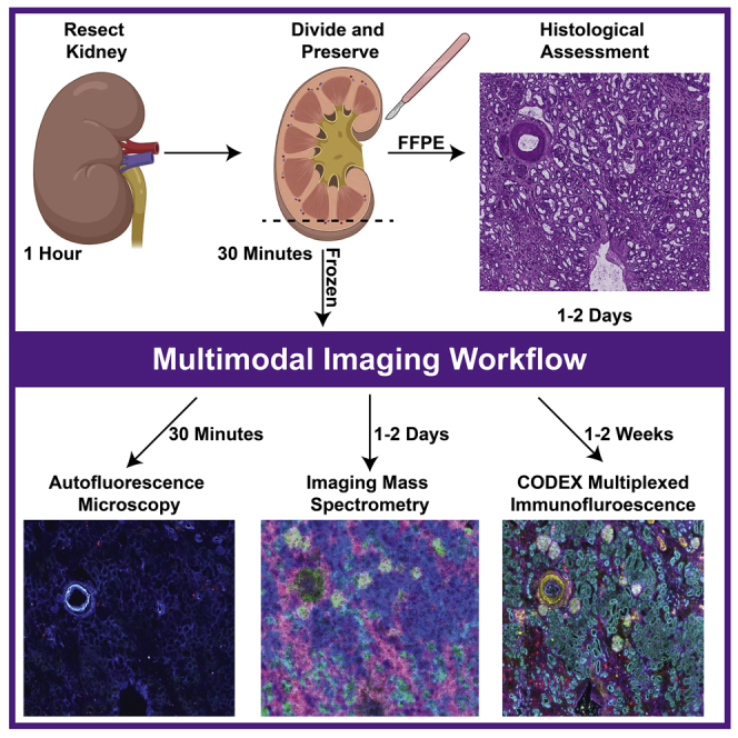
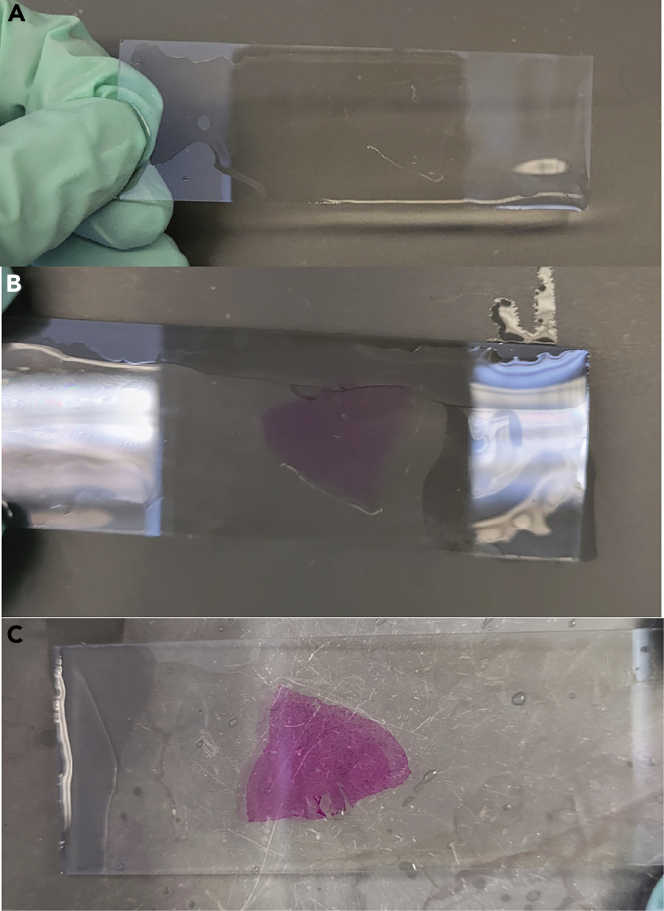
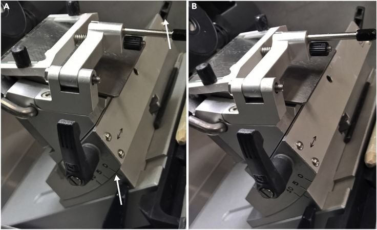
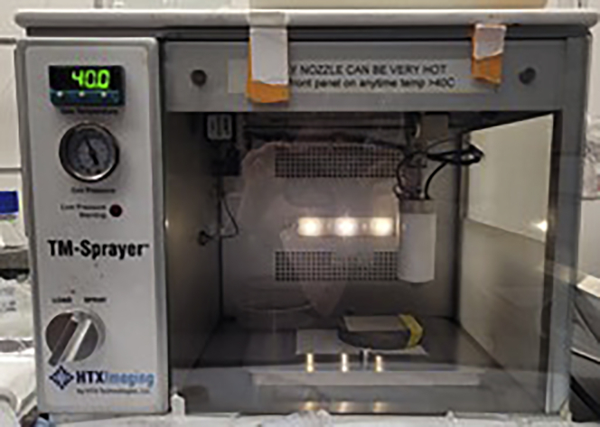
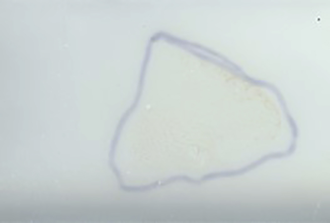
Here, we describe the preservation and preparation of human kidney tissue for interrogation by histopathology, imaging mass spectrometry, and multiplexed immunofluorescence. Custom image registration and integration techniques are used to create cellular and molecular atlases of this organ system. Through careful optimization, we ensure high-quality and reproducible datasets suitable for cross-patient comparisons that are essential to understanding human health and disease. Moreover, each of these steps can be adapted to other organ systems or diseases, enabling additional atlas efforts.

Keywords: Antibody; Chemistry; Health Sciences; Mass Spectrometry; Metabolomics; Microscopy.


Conflict of interest statement

The authors declare no competing interests.

Figures

None
Graphical abstract
Figure 1
Figure 1
Kidney resections Section of resected kidney with the superior and inferior poles labeled. (A) Whole kidney cut in half, coronally with superior and inferior poles labeled (black dotted line). (B) A portion of the kidney (∼80 × 60 × 4 mm) that is farthest away from the tumor (green dotted line). (C) The extracted portion is divided into thirds or fourths to ensure event freezing (blue dotted line).
Figure 2
Figure 2
Measurement guide Measure and record the length, width, and thickness of the kidney tissue pieces using gridded paper (as shown above) or other instruments.
Figure 3
Figure 3
Kidney embedding and freezing (A–D) (A) Label mold such that orientation of the kidney can be maintained. Images of unfrozen tissue (B), partially frozen tissue and CMC (C), and CMC embedded tissue ready for storage (D).
Figure 4
Figure 4
Schiff’s reagent stain example (A) Tissue after incubation with periodic acid. The tissue is clear and uncolored. (B) Tissue appearance after incubation with Schiff’s reagent. The tissue is light pink. (C) Tissue appearance after exposure to warm water. The tissue turns bright pink when exposed to warm water.
Figure 5
Figure 5
Cryostat configuration (A and B) (A) Unadjusted angle and (B) adjusted angle of the cryostat blade holder.
Figure 6
Figure 6
Matrix application configuration TM Sprayer set up for high resolution matrix coating.
Figure 7
Figure 7
Example of a matrix coated slide An example of uniform matrix coating of sample of a sample outlined using sharpie on the back of the slide.
Figure 8
Figure 8
Multimodal images from the same kidney tissue section Example images of a human kidney after following each step within the protocol: (A) PAS histological stain (B) autofluorescence microscopy image, (C) MALDI IMS of [SM(d34:1)+Na]+ (green), [PC(32:0)+H]+ (pink), and [PC(36:4)+H]+ (blue), (D) and CODEX IF of aquaporin 1 (teal), aquaporin 2 (yellow), uromodulin (red), and vimentin (purple). Scale bars are 500 nm.

References

    1. Caprioli R.M., Farmer T.B., Gile J. Molecular imaging of biological samples: localization of peptides and proteins using maldi-tof ms. Anal. Chem. 1997;69:4751–4760. - PubMed
    1. Fahy E., Subramaniam S., Murphy R.C., Nishijima M., Raetz C.R.H., Shimizu T., Spener F., Van Meer G., Wakelam M.J.O., Dennis E.A. Update of the lipid maps comprehensive classification system for lipids. J. Lipid Res. 2009;50:s9–s14. - PMC - PubMed
    1. Fahy E., Sud M., Cotter D., Subramaniam S. Lipid maps online tools for lipid research. Nucleic Acids Res. 2007;35:w606–w612. - PMC - PubMed
    1. Gill E.L., Yost R.A., Vedam-Mai V., Garrett T.J. Precast gelatin-based molds for tissue embedding compatible with mass spectrometry imaging. Anal. Chem. 2017;89:576–580. - PMC - PubMed
    1. Goltsev Y., Samusik N., Kennedy-Darling J., Bhate S., Hale M., Vazquez G., Black S., Nolan G.P. Deep profiling of mouse splenic architecture with codex multiplexed imaging. Cell. 2018;174:968–981.e15. - PMC - PubMed

Publication types