Review
. 2021 Sep 1;36(5):272-291.
doi: 10.1152/physiol.00004.2021.

Halogen-Induced Chemical Injury to the Mammalian Cardiopulmonary Systems

Affiliations
Review

Halogen-Induced Chemical Injury to the Mammalian Cardiopulmonary Systems

Dylan R Addis et al. Physiology (Bethesda). .

Abstract

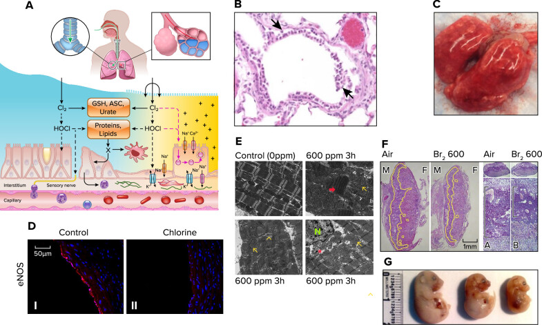
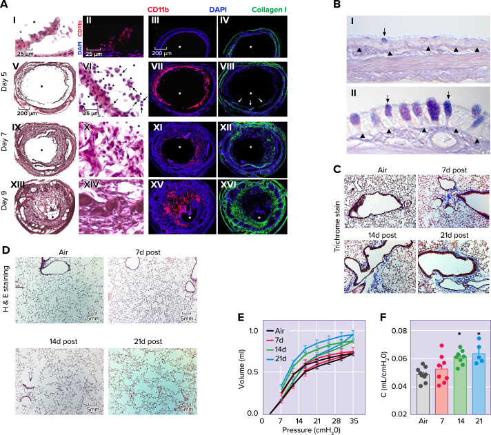
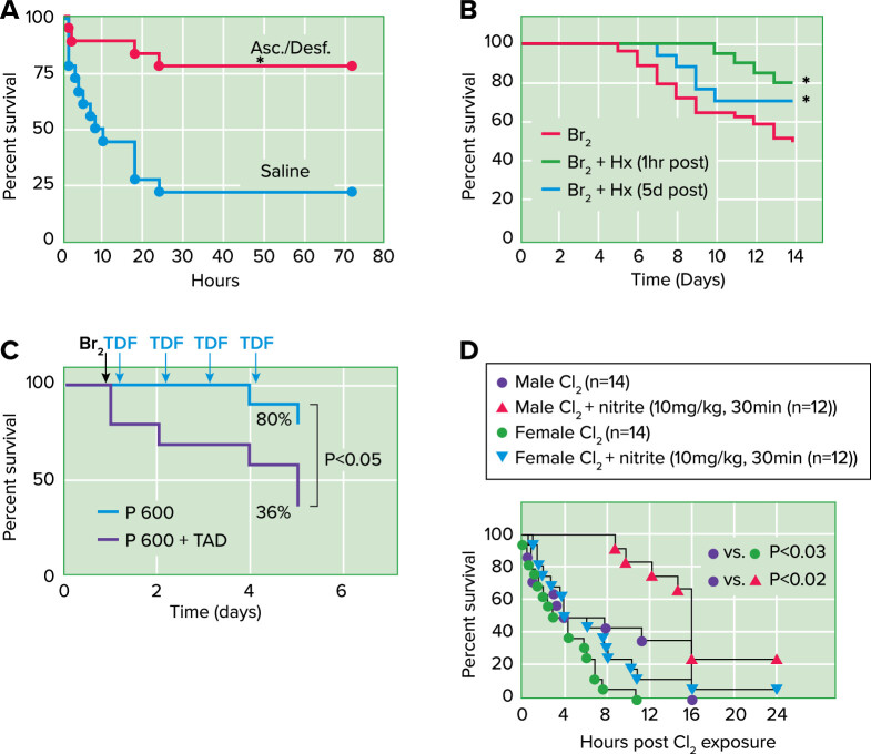
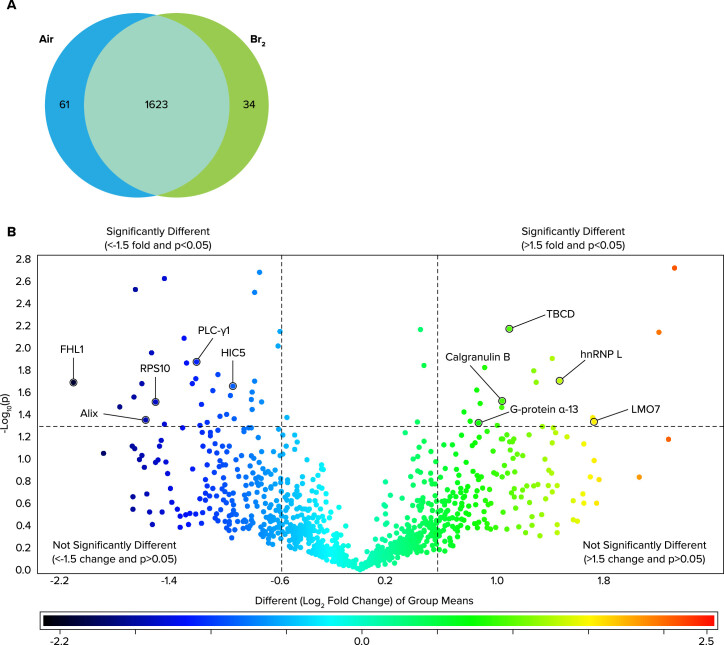
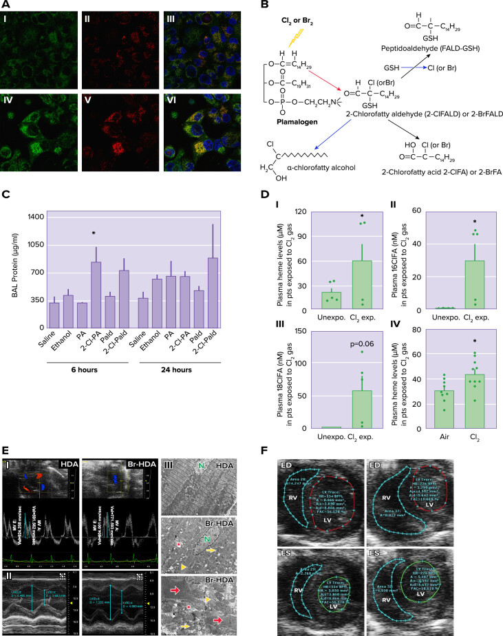
The halogens chlorine (Cl2) and bromine (Br2) are highly reactive oxidizing elements with widespread industrial applications and a history of development and use as chemical weapons. When inhaled, depending on the dose and duration of exposure, they cause acute and chronic injury to both the lungs and systemic organs that may result in the development of chronic changes (such as fibrosis) and death from cardiopulmonary failure. A number of conditions, such as viral infections, coexposure to other toxic gases, and pregnancy increase susceptibility to halogens significantly. Herein we review their danger to public health, their mechanisms of action, and the development of pharmacological agents that when administered post-exposure decrease morbidity and mortality.

Keywords: ARDS; antioxidants; bromine; chlorine; heme.


Figures

FIGURE 1.
FIGURE 1.
Halogen (Cl2 and Br2)-induced acute lung and systemic injury A: schema showing interaction of halogens with various components of the epithelial lining fluid and lung epithelial cells. At higher concentrations (>50 ppm), inhaled halogens reach the distal lung regions (from Ref. , with permission from the publisher). ASC, ascorbate; GSH, reduced glutathione. B: extensive denudation of airway epithelium in a rat exposed to Cl2 (400 ppm for 30 min) and returned to room air for 24 h. Lung sections were stained with hematoxylin-eosin. C: lungs of a mouse exposed to Cl2 (600 ppm and 45 min) and returned to room air for 24 h. There was significant hemorrhage and alveolar spaces were filled with protein-rich edema. D: Cl2 decreases endothelial nitric oxide synthase (eNOS) protein expression. I and II: representative immunofluorescence images for eNOS staining from aorta collected 24 h after exposure to air (I) or Cl2 (400 ppm, 30 min) (II). Red: eNOS; blue: Hoechst staining for nuclei (modified from Ref. , with permission from the publisher). E: bromine (Br2) inhalation causes disruption of the cardiac cytoskeleton and loss of the normal highly organized linear mitochondrial sarcomere integrity. Representative transmission electron microscopy (×3,200) images demonstrating a normal control heart (top left). The remaining 3 images demonstrate myofibrillar loss, contraction band necrosis (red arrow), loss of I bands, and disruption of z-disks (yellow arrowheads) in the left ventricle 3 h after Br2 exposure in addition to mitochondrial swelling (yellow arrows) and cristae lysis (red asterisk) [nucleus (N)] (modified from Ref. , with permission from the publisher). F: pregnant [embryonic day 14.5 (E14.5)] mice were exposed to air or 600 ppm Br2 for 30 min, returned to room. Representative hematoxylin-eosin-stained placenta sections at E18.5 with the junctional zone demarcated with yellow highlighting (left) as well as periodic acid-Schiff staining (right) of Br2-exposed pregnant mice revealed a reduced junctional zone (black bars) at E18.5 (modified from Ref. , with permission from the publisher). M, male; F, female; A, air; B, bromide. G: pregnant mice were exposed to air or Br2 (400 or 600 ppm for 30 min) at E14.5 and were returned to room air. Fetuses were delivered by C-section on E18.5. Notice marked fetal growth restriction that was dependent on Br2 concentration (modified from Ref. 55).
FIGURE 2.
FIGURE 2.
Halogen (Cl2 and Br2) induced chronic lung injury A: loss of epithelial integrity results in inflammatory cell infiltration, mesenchyme infiltration, and collagen deposition. I: hematoxylin-eosin staining of tracheal epithelium, 2 days after exposure to Cl2 (350 ppm for 30 min). II: immunofluorescent staining for CD11b (red) in a similar day 2 tissue section. In I and II, scale bars = 25 μm. Immunofluorescent staining of either CD11b (red) or collagen I (green) in a control tracheal section and sections harvested on days 5, 7, and 9 after high-dose Cl2 and CD11b (III, VII, XI, and XV, respectively) and collagen I (IV, VIII, XII, and XVI, respectively; scale bar, = 200 μm). In VII, arrows denote collagen 1+ cells infiltrating the lumen. Hematoxylin-and-eosin staining of tracheal sections harvested on days 5, 7, and 9 after high-dose Cl2 (V, IX, and XII, respectively; scale bar, = 200 μm); magnified images are presented VI, X, and XIV, respectively (scale bar, = 25 μm) In VI, arrowheads denote macrophages, whereas arrows denote neutrophils. All tissue sections are representative of 5–6 tracheas at each time. Asterisks denote the lumen (from Ref. , with permission from the publisher). B: histopathological effects of Cl2 inhalation in proximal airways, 7 days after exposure. Rats were exposed to air or 400 ppm Cl2 for 30 min. Tissue was collected 7 days after exposure, embedded in paraffin, and stained with Alcian blue and period acid-Schiff stain. I and II: representative light micrographs are of proximal airways from air-exposed (I) or 400 ppm Cl2 for 30 min (II). Arrows, mucous cells; arrowheads, basal lamina. Magnification bar = 50 μm (modified from Ref. , with permission from the publisher). C and D: male C57BL/6 mice were exposed to air or Br2 gas (400 ppm, 30 min) and returned to room air: peripheral lung tissue with Masson’s trichrome (C) or hematoxylin-eosin stain (H&E; D); notice increased accumulation collagen deposition (blue stain) primarily around airways on days 14 and 21 after Br2 (C) and marked enlargement of alveolar spaces (modified from Ref. , with permission from the publisher). E and F: quasistatic inflation and deflation pressure volume lung relationships (measured by flexiVent) (E) and lung compliance (F), measured as the slope of the deflation pressure-volume curve of mice exposed to Br2 gas (400 ppm, 30 min) and returned to room air at the indicated time points (modified from Ref. 68).
FIGURE 3.
FIGURE 3.
Countermeasures against halogen toxicity. Post-Cl2 administration of antioxidants increase survival Mice were exposed to 600 ppm of Cl2 for 45 min and returned to room air. A: they received intramuscular injections of ascorbate (Asc; 2 mg) and deferoxamine (Desf; 0.3 mg) in saline starting at 1 h after exposure and every 12 h thereafter up to 60 h after exposure. They also received aerosols of ascorbate (150 mg/mL) and deferoxamine (0.3577 mg/mL) at 1.5, 24, and 48 h after exposure in sterile water. Control mice received vehicle (saline for intramuscular injections; sterile water for aerosols) instead of antioxidants using identical protocols. Data points were fitted with Kaplan-Meier survival curves and compared with the log-rank test (P = 0.0007) (from Ref. 41). B: post-Br2 exposure administration of hemopexin increases survival. C57BL/6 mice were exposed to Br2 (400 ppm for 30 min) and returned to room air. At either 1 h or 5 days post-exposure they were given a single intraperitoneal injection of purified human hemopexin (Hx) (4 µg/g body wt) or vehicle. Survival was assessed in the next 14 days. The Kaplan-Meier curve demonstrated that Hx reduced mortality after Br2 exposure, even when was given 5 days later. *P < 0.05 vs. Br2 + saline by one-way ANOVA followed by Tukey’s post hoc test (68). C: post-Br2 administration of tadalafil (Cialis; TDF or TAD) decreases mortality of pregnant (P) mice. Nonpregnant (NP) and P gestational day 14.5 (E14.5) mice were exposed to air or to Br2 at 600 ppm for 30 min and returned to room air; they received tadalafil (TAD; 2 mg/kg body weight in 0.1 mL of sterile saline) or vehicle via oral gavage at 1 h post-exposure and every 24 h thereafter. Data show Kaplan-Meyer curves of P and NP mice with tadalafil or vehicle, post-Br2 exposure. NP mice exposed to Br2 and returned to room air lived longer than similarly exposed P mice (*P < 0.05) (from Ref. 55). D: post-Cl2 administration of nitrite improves survival. Male or female mice were exposed to Cl2 at 600 ppm, 45 min and then brought back to room air, and nitrite was administered by intramuscular injection 30 min post-exposure. Data show Kaplan-Meier survival curves. P < 0.03 between male and female Cl2-alone groups; P < 0.02 for nitrite therapy in males and P = 0.09 for nitrite therapy in females.
FIGURE 4.
FIGURE 4.
Global lung protein changes 24 h post-Br2 exposure. Adult male C57BL/6 mice, 8–10 wk old, were exposed to Br2 gas (600 ppm for 30 min) or air in environmental chambers and returned to room air Twenty-four hours later, their lungs were removed and proteins were processed for global proteomics analysis. A: Venn diagram demonstrating the total number of proteins identified across both groups in addition to those proteins found to be significantly changed following exposure to Br2 (increased vs. the other group). B: volcano plot of the log10(P) value vs. log2 fold change (Br2/air) demonstrating the distribution of the entire data set of proteins with upper limits (above the line) indicating statistically significant changes and outer limits (to the right and left of each line) indicating significant fold changes as outlined in materials and methods under statistics. Note that although fold change is visualized as log2, the cutoff value of ±1.5 was applied to the fold change before logging, thereby yielding the indicated ±0.6 limits. Various proteins that play a role in vascular permeability are identified by the arrows (from Ref. 84).
FIGURE 5.
FIGURE 5.
Production of secondary mediators A: exposure to Cl2 increases mitochondrial superoxide/hydrogen peroxide. I–VI: H441 cells immersed in 50 µl of artificial epithelial lining fluid were exposed to 95% air-5% CO2 as control (IIII) or Cl2 (100 ppm for 15 min) (IVVI) and returned to 95% air-5% CO2 for 1 h. Before imaging with confocal laser microscopy, the cells were incubated with MitoSOX (red) for 15 min and MitoTracker (green). II and V: most of the Cl2-exposed H441 cells showed higher levels of red fluorescence compared with controls. VI: MitoSOX fluorescence localized with MitoTracker (yellow), consistent with the presence of reactive species in mitochondria. Nuclei were counterstained with DAPI (blue). Quantitative analysis showed increased production of reactive intermediates at 6 h post-exposure (modified from Ref. , with permission from the publisher). B: plasmalogen-derived Cl2 and Br2 oxidation products. The vinyl ether bond of plasmalogens is targeted by Cl2, Br2, HOCl, and HOBr resulting in 2-chlorofatty(bromo)aldehyde production including 2-Cl(Br)-Pald and 2-Cl(Br)-Sald. The 2-chloro(bromo)fatty aldehydes are either oxidized to the 2-chloro(bromo)fatty acids and 2-Cl(Br)-PA and 2-Cl(Br)l-SA or reduced to the 2-chloro(bromo)fatty alcohols, 2-chloro(bromo)palmitoyl alcohol, and 2-chloro(bromo)stearoyl alcohol. Alternatively, nucleophilic attack of 2-chloro(bromo)fatty aldehydes by GSH results in either palmitaldehyde or stearaldehyde GSH adduct formation. R1 = C14H29 or C16H33 (modified from Ref. 147). C: effects of Cl-lipids on lung permeability and inflammation. C57bl/6 male mice were exposed to saline, ethanol, palmitate (PA), palmitic aldehyde (Pald), 2-Cl-PA, or 2-Cl-Pald by intranasal administration and lung injury was assessed by measuring total protein in the bronchoalveolar lavage levels of protein. Data are means ± SE (n = 4–6). *P < 0.05 relative to corresponding native fatty acid (from Ref. 147). BAL, bronchoalveolar lavage. D: plasma cell-free heme (CFH) and chlorinated lipids are elevated in humans and mice exposed to Cl2 gas. Blood was collected from patients (pts) exposed to Cl2 gas in the emergency room of the University of Alabama at Birmingham, 3–4 h post-exposure, stored for 72 h at 4°C at which time it was analyzed. Blood from human volunteers as controls was treated in the same fashion. I: plasma CFH levels in persons exposed to Cl2 were higher than the age- and sex-matched human controls. II and III: Cl2-exposed individuals also had elevated levels of 16ClFA (n = 4–5) (II) and 18ClFA (III). Similarly, adult male C57BL/6 mice exposed to Cl2 gas (400 ppm, 30 min) had increased levels of heme in plasma 24 h post-exposure. Individual values and means ± SE. *P < 0.05 vs. unexposed humans or air exposed mice; by unpaired t test (from Ref. 74). E: cardiac dysfunction and cardiomyocyte death with 2-bromohexadecanal (Br-HDA). I: left ventricular (LV) diastolic dysfunction was reflected by the decrease in mitral valve (MV) early and late wave velocities 4 h after injection of Br-HDA into the LV cavity. II: Br-HDA caused an increase in LV end-diastolic dimension and end-systolic dimension, resulting in a decrease in fractional shortening. Image demonstrates M-mode echocardiography of the LV before and 4 h after intracardiac injection of Br-HDA. III: Br-HDA caused extensive disruption of the cardiac cytoskeleton and loss of the normal highly organized linear mitochondrial sarcomere integrity. Transmission electron microscopy (×3,200 at top and middle and ×8,000 at bottom) demonstrated contraction band necrosis (red arrows), loss of I bands, and disruption of z-disk (yellow arrowheads) in the LV of rats administered Br-HDA as well as mitochondrial swelling (yellow arrows) and cristae lysis (red asterisk) (from Ref. 54). F: Br2-exposed pregnant mice exhibit diminished cardiac function at gestational day 18.5 (E18.5). Nonpregnant and pregnant (E14.5) mice were exposed to air or 600 ppm Br2 for 30 mi and returned to room air. Representative echosonography left ventricular (LV) and right ventricular (RV) traces at E18.5 of pregnant mice exposed to air (left) or Br2 with demarcation of ventricular sizes (ED, end diastole; ES, end systole) (from Ref. 55).

Similar articles

Cited by

References

    1. Summerhill EM, Hoyle GW, Jordt SE, Jugg BJ, Martin JG, Matalon S, Patterson SE, Prezant DJ, Sciuto AM, Svendsen ER, White CW, Veress LA, ATS Terrorism and Inhalational Disasters Section of the Environmental, Occupational, and Population Health Assembly. An Official American Thoracic Society Workshop Report: Chemical Inhalational Disasters. Biology of Lung Injury, Development of Novel Therapeutics, and Medical Preparedness. Ann Am Thorac Soc 14: 1060–1072, 2017. doi:10.1513/AnnalsATS.201704-297WS. - DOI - PMC - PubMed
    1. Carlisle M, Lam A, Svendsen ER, Aggarwal S, Matalon S. Chlorine-induced cardiopulmonary injury. Ann N Y Acad Sci 1374: 159–167, 2016. doi:10.1111/nyas.13091. - DOI - PMC - PubMed
    1. Hoyle GW, Svendsen ER. Persistent effects of chlorine inhalation on respiratory health. Ann N Y Acad Sci 1378: 33–40, 2016. doi:10.1111/nyas.13139. - DOI - PMC - PubMed
    1. Clark KA, Karmaus WJ, Mohr LC, Cai B, Balte P, Gibson JJ, Ownby D, Lawson AB, Vena JE, Svendsen ER. Lung function before and after a large chlorine gas release in Graniteville, South Carolina. Ann Am Thorac Soc 13: 356–363, 2016. doi:10.1513/AnnalsATS.201508-525OC. - DOI - PMC - PubMed
    1. Addis DR, Lambert JA, Ford DA, Jilling T, Matalon S. Halogen gas exposure: toxic effects on the parturient. Toxicol Mech Methods 31: 272–287, 2020. doi:10.1080/15376516.2020.1736702. - DOI - PMC - PubMed

Publication types

LinkOut - more resources