Review
. 2021 Oct;103(9):e278-e281.
doi: 10.1308/rcsann.2020.7134. Epub 2021 Aug 25.

Chronic sciatic nerve compression secondary to arteriovenous malformation: case discussion and literature review

Affiliations
Review

Chronic sciatic nerve compression secondary to arteriovenous malformation: case discussion and literature review

K S Johal et al. Ann R Coll Surg Engl. 2021 Oct.

Abstract

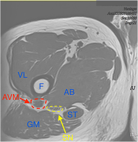
Sciatic nerve symptomatology may arise from both intra- and extra-neural pathology, at any point along descent from the sacral plexus to its bifurcation. The potential aetiology is broad, ranging from degenerative spinal disease to muscle, bony and vascular pathology. We present an extremely unusual case of position and exercise-induced nerve compression secondary to arteriovenous malformation and review the potential extraspinal causes, many of which may be ameliorated by surgical excision or decompression. We further discuss the usefulness of diagnostic imaging, specific clinical tests and histopathological tools that may aid in management.

Keywords: AVM; Compression; Sciatic nerve; Sciatica.


Figures

Figure 1
Figure 1
Sagittal cross-section MRI at sub-trochanteric level, depicting lesion (red arrow) in posterior right thigh. Inset: Close-up of lesion with feeding vessels shown.
Figure 2
Figure 2
Relations of lesion to sciatic nerve and muscle groups on sagittal MRI. (AB = adductor brevis, AVM = arteriovenous malformation, GM = gluteus maximus, SN = sciatic nerve, ST = semitendinosus, VL = vastus lateralis).
Figure 3
Figure 3
Intra-operative picture. Sciatic nerve (yellow dotted line) shown running over inferior edge of AVM (green dotted line). Inset: With sciatic nerve retracted extent of AVM shown, marked by artery forceps.
Figure 4
Figure 4
Histology demonstrating thick muscle layer and elastin staining of a large calibre artery, in keeping with diagnosis of AVM.

References

    1. Ropper AH, Zafonte RD. Sciatica. N Engl J Med 2015; 372: 1240–1248.10.1056/NEJMra1410151 - DOI - PubMed
    1. Porchet F, Wietlisbach V, Burnand Bet al. . Relationship between severity of lumbar disc disease and disability scores in sciatica patients. Neurosurgery 2002; 50: 1253–1260.10.1097/00006123-200206000-00014 - DOI - PubMed
    1. Hopayian K, Song F, Riera R, Sambandan S. The clinical features of the piriformis syndrome: a systematic review. Eur Spine J 2010; 19: 2095–2109.10.1007/s00586-010-1504-9 - DOI - PMC - PubMed
    1. Ilica AT, Yasar E, Sanal HTet al. . Sciatic nerve compression due to femoral neck osteochondroma: MDCT and MR findings. Clin Rheumatol 2008; 27: 403–404.10.1007/s10067-007-0761-4 - DOI - PubMed
    1. Reinstein L, Eckholdt JW. Sciatic nerve compression by preexisting heterotopic ossification during general anesthesia in the dorsal lithotomy position. Arch Phys Med Rehabil 1983; 64: 65–68. - PubMed