. 2021 Sep 3;9(1):e0102321.
doi: 10.1128/Spectrum.01023-21. Epub 2021 Aug 25.

Mechanistic Insights into the Capsule-Targeting Depolymerase from a Klebsiella pneumoniae Bacteriophage

Affiliations

Mechanistic Insights into the Capsule-Targeting Depolymerase from a Klebsiella pneumoniae Bacteriophage

Rhys A Dunstan et al. Microbiol Spectr. .

Abstract

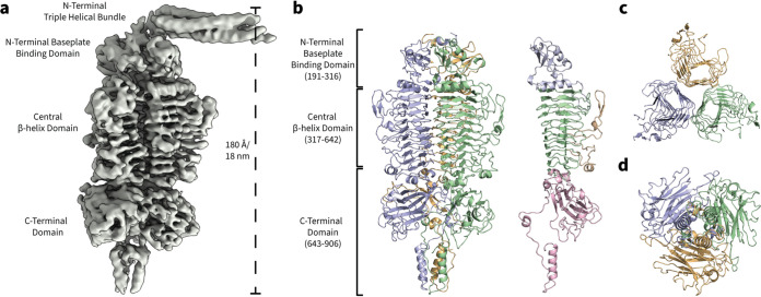
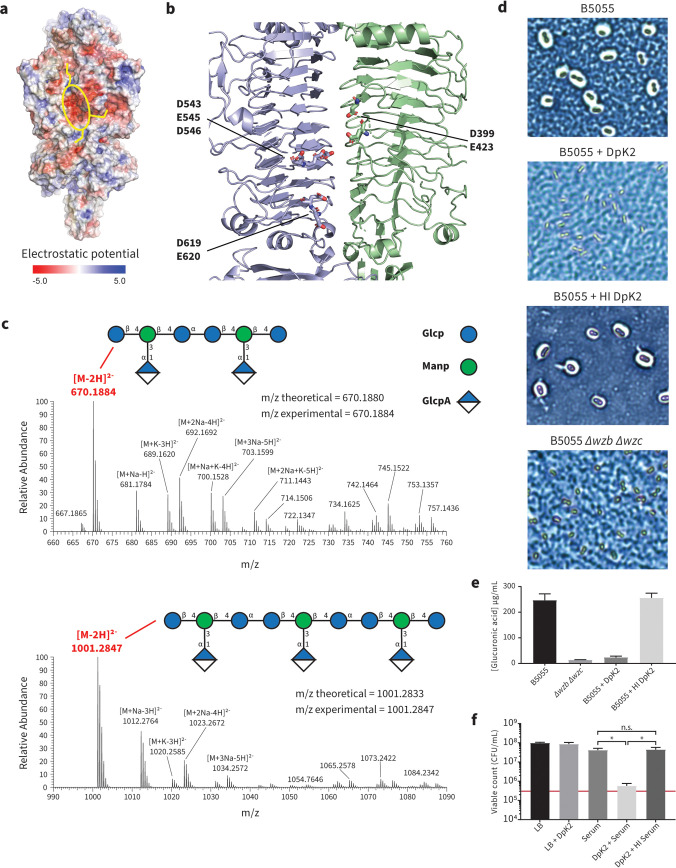
The production of capsular polysaccharides by Klebsiella pneumoniae protects the bacterial cell from harmful environmental factors such as antimicrobial compounds and infection by bacteriophages (phages). To bypass this protective barrier, some phages encode polysaccharide-degrading enzymes referred to as depolymerases to provide access to cell surface receptors. Here, we characterized the phage RAD2, which infects K. pneumoniae strains that produce the widespread, hypervirulence-associated K2-type capsular polysaccharide. Using transposon-directed insertion sequencing, we have shown that the production of capsule is an absolute requirement for efficient RAD2 infection by serving as a first-stage receptor. We have identified the depolymerase responsible for recognition and degradation of the capsule, determined that the depolymerase forms globular appendages on the phage virion tail tip, and present the cryo-electron microscopy structure of the RAD2 capsule depolymerase at 2.7-Å resolution. A putative active site for the enzyme was identified, comprising clustered negatively charged residues that could facilitate the hydrolysis of target polysaccharides. Enzymatic assays coupled with mass spectrometric analyses of digested oligosaccharide products provided further mechanistic insight into the hydrolase activity of the enzyme, which, when incubated with K. pneumoniae, removes the capsule and sensitizes the cells to serum-induced killing. Overall, these findings expand our understanding of how phages target the Klebsiella capsule for infection, providing a framework for the use of depolymerases as antivirulence agents against this medically important pathogen. IMPORTANCE Klebsiella pneumoniae is a medically important pathogen that produces a thick protective capsule that is essential for pathogenicity. Phages are natural predators of bacteria, and many encode diverse "capsule depolymerases" which specifically degrade the capsule of their hosts, an exploitable trait for potential therapies. We have determined the first structure of a depolymerase that targets the clinically relevant K2 capsule and have identified its putative active site, providing hints to its mechanism of action. We also show that Klebsiella cells treated with a recombinant form of the depolymerase are stripped of capsule, inhibiting their ability to grow in the presence of serum, demonstrating the anti-infective potential of these robust and readily producible enzymes against encapsulated bacterial pathogens such as K. pneumoniae.

Keywords: Klebsiella; bacteriophages; capsular polysaccharide; cryo-EM; depolymerase.


Figures

FIG 1
FIG 1
Isolation and characterization of Klebsiella phage RAD2. (a) Plaque morphology of RAD2 after infection of K. pneumoniae B5055. Scale bar, 10 mm. (b) Samples of phage RAD2 were purified using cesium chloride gradients and subjected to TEM analysis. White arrows highlight the globular tail appendages. Scale bar, 50 nm. (c) Circular representation of the RAD2 genome with putative functional assignment of the predicted open reading frames based on sequence analysis using BLASTp, HMMER, and HHpred color-coded by predicted function (pink, terminase; blue, DNA modification; yellow, nuclease; green, host cell lysis; red, structural or virion assembly; gray, unknown). (d) Representative images of cesium chloride-purified samples of RAD2 analyzed by immunogold labeling with antibodies raised against DpK2. Scale bar, 50 nm.
FIG 2
FIG 2
The K2 capsule is required for Klebsiella phage RAD2 infection. Summary of the TraDIS screen results. Transposon insertion sites mapped onto genes involved in CPS biosynthesis, export, and regulation. (a to c) An enrichment of insertions was observed in many genes (highlighted in red) of the CPS locus (a), the transcriptional antiterminator rfaH (b), and the UTP-glucose-1-phosphate uridylyltransferase galU (c) after infection by RAD2 compared to the uninfected input library. Scale bar, 1.0 kb. (d) Spot assays of diluted RAD2 preparations onto top agar layers containing K. pneumoniae B5055 wild type, B5055 Δwzb Δwzc, or the complemented B5055 Δwzb Δwzc mutant expressing plasmid-encoded Wzb and Wzc from an anhydrotetracycline-inducible promoter. The neat RAD2 titer used to prepare the serially diluted samples was ∼108 PFU/ml.
FIG 3
FIG 3
Structure of the tailspike protein DpK2 from Klebsiella phage RAD2. (a) Single-particle cryo-EM electron density of DpK2 refined using C1 symmetry. (b) High-resolution structure of the DpK2 trimer. The individual protein monomers are colored in green, blue, and yellow. Structural domains are highlighted on the Dpk2 monomer: N-terminal baseplate binding domain (residues 191 to 316, blue), the central β-helix domain (residues 317 to 642, green) with extended outward facing loop structures (yellow), and a C-terminal domain (residues 643 to 906, pink). (c) Cutaway top-down view of the three adjacent β-helices of the DpK2 trimer. Each helix is formed by an individual monomer assembled along the length of the protein. (d) Bottom-up view of the DpK2 trimer showing the C-terminal domain, which is formed predominantly by three individual β-sandwich folds and a short α-helical bundle forming a spike-like structure.
FIG 4
FIG 4
Recombinant DpK2 is an active enzyme. (a) Halo spot assays measuring the activity of DpK2 using double overlay plates. The top agar was preinoculated with K. pneumoniae B5055 before spotting it with increasing amounts of purified DpK2 (5 to 500 ng) or TBS. (b) Extracts of purified K2 capsule treated with either 1 μg of DpK2 or TBS analyzed by 3-to-14% SDS-PAGE and stained with Alcian blue. (c) Thermal stability of DpK2 measured with the colorimetric compound pHBAH. DpK2 (500 μg) was preincubated at the indicated temperatures for 30 min prior to the addition of purified K2 capsule. Absorbance readings at 420 nm were used to measure the generation of reducing sugars produced by the cleavage of capsular polysaccharide. Error bars represent standard deviations of the results of three biological replicates.
FIG 5
FIG 5
Mechanistic insight into the enzymatic activity of DpK2. (a) The electrostatic potential (negative, red; positive, blue) mapped onto the surface of the DpK2 trimer, highlighting a strong negatively charged cavity (yellow oval) connected to adjacent grooves (yellow lines). (b) The negatively charged cavity is formed within an interchain groove between neighboring DpK2 monomers. (c) Negative-mode mass spectra of DpK2-treated capsule analyzed by LC-MS. The prominent masses are highlighted in red. The multiple masses observed within these spectra can be attributed to the formation of ion adducts that differ in the nature of ionizing species. The cartoon structure of the K2 repeating units is depicted with the theoretical and experimental m/z ratios highlighted for both cleavage products. Glc, glucose; Man, mannose; GlcA, glucuronic acid; p, sugars in pyranose form. (d) Representative images of enzyme-treated K. pneumoniae cells visualized by Maneval’s staining and light microscopy. (e) Quantification of cell-associated uronic acid (μg/ml). Capsular polysaccharides were isolated from whole cells using phenol-water extraction after treatment with active or heat-inactivated DpK2. Samples were hydrolyzed in sulfuric acid prior to the addition of 3-hydroxyldiphenol and subsequent absorbance reading at 520 nm. The uronic acid concentration was measured using a defined glucuronic acid standard curve. Error bars represent standard deviations of the results of three biological duplicates. (f) Quantitation of K. pneumoniae B5055 growth in the presence of DpK2 and serum as measured by the number of recovered CFU/ml. Cells were treated with either DpK2, serum, DpK2 plus serum, or DpK2 plus heat-inactivated (HI) serum for 3 h. The red bar represents the number of CFU/ml of the starting inoculum prior to the addition of serum. Overall statistical significance was determined by one-way analysis of variance (ANOVA) (P value < 0.0001 for all conditions). Further statistical analyses were performed using Turkey’s multiple-comparison test for all serum treatments (*, P < 0.0001; n.s., not significant). Error bars represent standard deviations of the results of three biological duplicates.

References

    1. Podschun R, Ullmann U. 1998. Klebsiella spp. as nosocomial pathogens: epidemiology, taxonomy, typing methods, and pathogenicity factors. Clin Microbiol Rev 11:589–603. doi: 10.1128/CMR.11.4.589. - DOI - PMC - PubMed
    1. Holt KE, Wertheim H, Zadoks RN, Baker S, Whitehouse CA, Dance D, Jenney A, Connor TR, Hsu LY, Severin J, Brisse S, Cao H, Wilksch J, Gorrie C, Schultz MB, Edwards DJ, Nguyen KV, Nguyen TV, Dao TT, Mensink M, Minh VL, Nhu NT, Schultsz C, Kuntaman K, Newton PN, Moore CE, Strugnell RA, Thomson NR. 2015. Genomic analysis of diversity, population structure, virulence, and antimicrobial resistance in Klebsiella pneumoniae, an urgent threat to public health. Proc Natl Acad Sci USA 112:E3574–E3581. doi: 10.1073/pnas.1501049112. - DOI - PMC - PubMed
    1. Paczosa MK, Mecsas J. 2016. Klebsiella pneumoniae: going on the offense with a strong defense. Microbiol Mol Biol Rev 80:629–661. doi: 10.1128/MMBR.00078-15. - DOI - PMC - PubMed
    1. Sahly H, Podschun R, Ullmann U. 2000. Klebsiella infections in the immunocompromised host. Adv Exp Med Biol 479:237–249. doi: 10.1007/0-306-46831-X_21. - DOI - PubMed
    1. Gomez-Simmonds A, Uhlemann AC. 2017. Clinical implications of genomic adaptation and evolution of carbapenem-resistant Klebsiella pneumoniae. J Infect Dis 215:S18–S27. doi: 10.1093/infdis/jiw378. - DOI - PMC - PubMed

Publication types

MeSH terms

LinkOut - more resources