Sebaceous adenoma of the conjunctiva and caruncle: a clinicopathological report of three cases and literature review
- PMID: 34431898
- PMCID: PMC11826563
- DOI: 10.5935/0004-2749.20220023
Sebaceous adenoma of the conjunctiva and caruncle: a clinicopathological report of three cases and literature review
Abstract
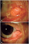
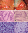
Sebaceous tumors of the conjunctiva and caruncle are rare conditions, accounting for 1% of caruncle lesions and even lower among conjunctival lesions. Almost 50% of cases are associated with Muir-Torre syndrome, a rare autosomal-dominant condition characterized by at least one sebaceous skin tumor and one visceral malignancy. We report 3 cases of sebaceous adenoma with different presentations that were submitted to excisional biopsy and immunohistochemical study. Diagnosis of these tumors should increase the level of suspicion and lead to clinical investigation to rule out neoplasms, particularly because in up to 41% of cases, these can be the first sign of the disease.
Tumores sebáceos da conjuntiva e carúncula são condições raras, sendo mais encontrados nas pálpebras. O adenoma sebáceo é responsável por 1% das lesões carunculares e ainda menos frequente nas lesões da conjuntiva. Esse diagnóstico é de suma importância pois quase 50% desses pacientes podem ser diagnosticados com a Síndrome de Muir-Torre, uma condição autossômica dominante rara que é caracterizada pela presença de ao menos um tumor de pele sebáceo e uma neoplasia visceral (gastrointestinal, câncer geniturinário e de mama). O estudo tem como objetivo relatar 3 casos de adenoma sebáceo com diferentes apresentações, que foram submetidos a biópsia excisional e estudo imuno-histoquímico. O diagnóstico desses tumores deve levantar suspeitas e aconselhar a investigação clínica para descartar outras neoplasias, principalmente porque em até 41% dos casos esse pode ser o primeiro sinal da Síndrome de Muir-Torre.
Conflict of interest statement
Figures


References
-
- Kaeser PF, Uffer S, Zografos L, Hamédani M. Tumors of the caruncle: a clinicopathologic correlation. Am J Ophthalmol. 2006;142(3):448–455. - PubMed
-
- Ilhan HD, Turkoglu EB, Bilgin AB, Bassorgun I, Dogan ME, Unal M. A unique case of isolated sebaceous adenoma of the bulbar conjunctiva. Arq Bras Oftalmol. 2016;79(4):253–254. - PubMed
-
- Tok L, Tok OY, Argun M, Ciris IM, Baspinar S, Gunes A. Corneal limbal sebaceous adenoma. Cornea. 2014;33(4):425–427. - PubMed
-
- Cohen PR, Kohn SR, Kurzrock R. Association of sebaceous gland tumors and internal malignancy: the Muir-Torre syndrome. Am J Med. 1991;90(5):606–613. - PubMed
-
- Luthra CL, Doxanas MT, Green WR. Lesions of the caruncle: a clinicohistopathologic study. Surv Ophthalmol. 1978;23(3):183–195. - PubMed
Publication types
MeSH terms
LinkOut - more resources
Full Text Sources

