. 2021 Sep 27;49(17):10166-10177.
doi: 10.1093/nar/gkab736.

BusR senses bipartite DNA binding motifs by a unique molecular ruler architecture

Affiliations

BusR senses bipartite DNA binding motifs by a unique molecular ruler architecture

Adrian M Bandera et al. Nucleic Acids Res. .

Abstract

The cyclic dinucleotide second messenger c-di-AMP is a major player in regulation of potassium homeostasis and osmolyte transport in a variety of bacteria. Along with various direct interactions with proteins such as potassium channels, the second messenger also specifically binds to transcription factors, thereby altering the processes in the cell on the transcriptional level. We here describe the structural and biochemical characterization of BusR from the human pathogen Streptococcus agalactiae. BusR is a member of a yet structurally uncharacterized subfamily of the GntR family of transcription factors that downregulates transcription of the genes for the BusA (OpuA) glycine-betaine transporter upon c-di-AMP binding. We report crystal structures of full-length BusR, its apo and c-di-AMP bound effector domain, as well as cryo-EM structures of BusR bound to its operator DNA. Our structural data, supported by biochemical and biophysical data, reveal that BusR utilizes a unique domain assembly with a tetrameric coiled-coil in between the binding platforms, serving as a molecular ruler to specifically recognize a 22 bp separated bipartite binding motif. Binding of c-di-AMP to BusR induces a shift in equilibrium from an inactivated towards an activated state that allows BusR to bind the target DNA, leading to transcriptional repression.


Figures

Graphical Abstract
Graphical Abstract
c-di-AMP mediated transcriptional control by BusR.
Figure 1.
Figure 1.
Crystal structure of apo S. agalactiae BusR and ligand binding. (A) Cartoon representation of the tetrameric crystal structure of apo BusR, colored by chain. The respective domains are labelled to illustrate the antiparallel head-to-tail arrangement. The ligand binding pocket is highlighted by a dashed circle. (B) Side view of the apo crystal structure, rotated by 90°. (C) Binding curve and fits of ITC measurements of BusR titrated with c-di-AMP (orange, KD= 112 ± 7 nM), 5′-pApA (brown, no binding) and c-di-GMP (blue, no binding) (n = 3). (D) Schematic overview of the secondary structure of a single BusR monomer from the apo crystal structure. The helix elongation of the central coiled-coils labelled αCC′ is only present in the light blue and light brown colored monomers (see panel A and B), while unstructured in the others.
Figure 2.
Figure 2.
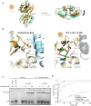
Release of autoinhibition upon c-di-AMP binding to BusR. (A) Front and side view of superimposed crystal structures of apo RCK_C domain (orange) and ligand bound RCK_C domain (cyan, c-di-AMP in dark green). Ligand induced movement of the monomers in respect to each other is indicated by a black arrow (1.31 Å rmsd for apo RCK_C compared to ligand bound RCK_C). (B) Close-up on RCK_C – wHTHinhib interface in the full-length (left) and c-di-AMP bound RCK_C (right) crystal structure. In the full-length apo state a tight interface is formed around residues Trp159 (RCK_C chain A, light brown), Pro179-Phe182 (RCK_C chain B, dark brown) and Tyr13 (wHTHinhib, blue). Binding of c-di-AMP induces rotational movement (black arrow) of the RCK_C monomers in respect to each other that would cause sterical clashes with helices αG1 and αG3 (grey and transparent), which frees the wHTHinhib domain for subsequent DNA binding. (C) EMSA experiments: c-di-AMP titrated to 100 nM wt BusR leads to increased affinity for operator DNA (20 nM), while related nucleotides show no effect. The mutation of Trp159Ala disturbs the RCK_C–wHTHinhib interface and thereby allows for DNA binding of BusR in absence of c-di-AMP (n = 3). (D) Steady-state affinity binding fits of SPR measurements using a single site binding model. Titration of BusR and BusR Trp159Ala to pAB1 in presence and absence of c-di-AMP (10 μM) (n = 3).
Figure 3.
Figure 3.
BusR bound to operator DNA. (A) cryoEM structure of BusR bound to operator DNA pAB depicted as side view (left), top view (top right) and front view (bottom right). The binding motifs are colored in purple. Large domain rearrangement occurs from apo to DNA bound BusR. The RCK_C–wHTHinhib interface is broken and all four wHTH motifs rotate backwards to face and bind the DNA. (B) The DNA binding domain shows great variability in its interaction with the DNA. The wHTHfree rather interacts with the phosphate backbone instead of the bases (left part, for reasons of better visibility wHTHinhib is not shown), whereas wHTHinhib is deeply buried in the major groove with its wing bound to the minor groove (right part, wHTHfree is not shown). (C) BusR distinguishes its native substrate (pAB1 or pAB2) from non-target DNA. Alterations to the DNA severely disrupt binding of BusR in EMSAs. Deletion of one (BS-A or BS-B) or both binding motifs (0BS) from pAB1 abolishes DNA binding in EMSAs. Also changes to the position of the binding motifs, either by elongation of the DNA in between the binding motifs by 5 base pairs (+5 bp) or by shortening of the distance in between by 10 base pairs (−10 bp) prevents BusR from recognizing its substrate (BusR = 100 nM, DNA = 20 nM, c-di-AMP = 10 μM, n = 3). (D) EMSA with the promoter region containing both pAB1 and pAB2. Two complex bands are observed upon titration of BusR. A control with only one binding site present results in the lower complex band, indicating that this is a BusR:DNA complex of 1:1 stochiometry and linear instead of looped DNA (20 nM DNA, 10 μM c-di-AMP; n = 3). (E) Schematic overview of the two binding sites of BusR within the promotor region of BusA. Included are the −35 and −10 transcriptional elements, the transcriptional start site (TSS) and the start codon (ATG). (F) Schematic representation of the domain rearrangement and mechanism. Two molecules of c-di-AMP bind to the RCK_C domains. Consequently, the adjacent wHTHinhib domain is released and can now freely rotate towards the DNA. Upon binding the DNA is bent by 18°.

References

    1. Witte G., Hartung S., Büttner K., Hopfner K.P.. Structural biochemistry of a bacterial checkpoint protein reveals diadenylate cyclase activity regulated by DNA recombination intermediates. Mol. Cell. 2008; 30:167–178. - PubMed
    1. Bejerano-Sagie M., Oppenheimer-Shaanan Y., Berlatzky I., Rouvinski A., Meyerovich M., Ben-Yehuda S.. A checkpoint protein that scans the chromosome for damage at the start of sporulation in Bacillus subtilis. Cell. 2006; 125:679–690. - PubMed
    1. Oppenheimer-Shaanan Y., Wexselblatt E., Katzhendler J., Yavin E., Ben-Yehuda S.. c-di-AMP reports DNA integrity during sporulation in Bacillus subtilis. EMBO Rep. 2011; 12:594–601. - PMC - PubMed
    1. Römling U. Great times for small molecules: c-di-AMP, a second messenger candidate in Bacteria and Archaea. Sci. Signal. 2008; 1:pe39. - PubMed
    1. Stülke J., Krüger L.. Cyclic di-AMP signaling in bacteria. Annu. Rev. Microbiol. 2020; 74:159–179. - PubMed

Publication types

MeSH terms