. 2021 Aug 25;32(9):108.
doi: 10.1007/s10856-021-06563-1.

Radio frequency plasma assisted surface modification of Fe3O4 nanoparticles using polyaniline/polypyrrole for bioimaging and magnetic hyperthermia applications

Affiliations

Radio frequency plasma assisted surface modification of Fe3O4 nanoparticles using polyaniline/polypyrrole for bioimaging and magnetic hyperthermia applications

Beena Mol et al. J Mater Sci Mater Med. .

Abstract

Surface modification of superparamagnetic Fe3O4 nanoparticles using polymers (polyaniline/polypyrrole) was done by radio frequency (r.f.) plasma polymerization technique and characterized by XRD, TEM, TG/DTA and VSM. Surface-passivated Fe3O4 nanoparticles with polymers were having spherical/rod-shaped structures with superparamagnetic properties. Broad visible photoluminescence emission bands were observed at 445 and 580 nm for polyaniline-coated Fe3O4 and at 488 nm for polypyrrole-coated Fe3O4. These samples exhibit good fluorescence emissions with L929 cellular assay and were non-toxic. Magnetic hyperthermia response of Fe3O4 and polymer (polyaniline/polypyrrole)-coated Fe3O4 was evaluated and all the samples exhibit hyperthermia activity in the range of 42-45 °C. Specific loss power (SLP) values of polyaniline and polypyrrole-coated Fe3O4 nanoparticles (5 and 10 mg/ml) exhibit a controlled heat generation with an increase in the magnetic field.


Conflict of interest statement

The authors declare no competing interests.

Figures

Fig. 1
Fig. 1
XRD pattern of a Fe3O4, b PANI-coated Fe3O4, and c polypyrrole-coated Fe3O4 nanoparticles
Fig. 2
Fig. 2
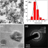
a TEM image of Fe3O4, b Histogram of particle sizes obtained from TEM, c HRTEM image and d SAED pattern of Fe3O4
Fig. 3
Fig. 3
a TEM image of iron oxide nanoparticles with spherical and rod-shaped morphology (Fe3O4). The inset shows the average diameter of nanorods and b HRTEM image of nanorod iron oxide
Fig. 4
Fig. 4
a TEM image of PANI-coated Fe3O4, b histogram of particle sizes obtained from TEM, c HRTEM image and d SAED pattern of PANI-coated Fe3O4
Fig. 5
Fig. 5
a TEM image of polypyrrole-coated Fe3O4, b histogram of particle sizes obtained from TEM, c HRTEM image and d SAED pattern of polypyrrole-coated Fe3O4
Fig. 6
Fig. 6
Photoluminescence spectra of Fe3O4, PANI-coated Fe3O4 and polypyrrole-coated Fe3O4 under an excitation wavelength of 325 nm
Fig. 7
Fig. 7
Bright field and fluorescence images of L929 cell lines treated with a Fe3O4, b polyaniline-coated Fe3O4 nanoparticles, and c polypyrrole-coated Fe3O4 nanoparticles (×40 magnification)
Fig. 8
Fig. 8
Cytotoxicity studies of MTT assays for different concentrations of Fe3O4, PANI and polypyrrole-coated Fe3O4 nanoparticles incubated in U87 cell lines
Fig. 9
Fig. 9
Magnetic hysteresis curves of a Fe3O4, b PANI-coated Fe3O4, and c polypyrrole-coated Fe3O4 at 300 K
Fig. 10
Fig. 10
Time-temperature graph of a Fe3O4, b PANI-coated Fe3O4, and c polypyrrole-coated Fe3O4 with 1 mg concentration on exposure to 200–400 A (ac) current with a frequency of 280 kHz
Fig. 11
Fig. 11
Time–temperature graph of a Fe3O4, b PANI-coated Fe3O4, and c polypyrrole-coated Fe3O4 of 3 mg concentration on exposure to 200–400 A (ac) current with a frequency of 280 kHz

References

    1. McBain SC, Yiu HH, Dobson J. Magnetic nanoparticles for gene and drug delivery. Int J Nanomed. 2008;3:169–180. - PMC - PubMed
    1. Wu W, Wu Z, Yu T, Jiang C, Kim WS. Recent progress on magnetic iron oxide nanoparticles: synthesis, surface functional strategies and biomedical applications. Sci Technol Adv Mater. 2015;16:023501. doi: 10.1088/1468-6996/16/2/023501. - DOI - PMC - PubMed
    1. Bao Y, Sherwood JA, Sun Z. Magnetic iron oxide nanoparticles as T1 contrast agents for magnetic resonance imaging. J Mater Chem C. 2018;6:1280–1290. doi: 10.1039/C7TC05854C. - DOI
    1. Silva AC, Oliveira TR, Mamani JB, Malheiros SMF, Malavolta L, Pavon LF, et al. Application of hyperthermia induced by superparamagnetic iron oxide nanoparticles in glioma treatment. Int J Nanomed. 2011;6:591–603. - PMC - PubMed
    1. Abenojar EC, Wickramasinghe S, Bas-Concepcion J, Samia ACS. Structural effects on the magnetic hyperthermia properties of iron oxide nanoparticles. Prog Nat Sci Mater. 2016;26:440–448. doi: 10.1016/j.pnsc.2016.09.004. - DOI

MeSH terms