. 2021 Aug 25;17(8):e1009839.
doi: 10.1371/journal.ppat.1009839. eCollection 2021 Aug.

Activation of Cph1 causes ß(1,3)-glucan unmasking in Candida albicans and attenuates virulence in mice in a neutrophil-dependent manner

Affiliations

Activation of Cph1 causes ß(1,3)-glucan unmasking in Candida albicans and attenuates virulence in mice in a neutrophil-dependent manner

Andrew S Wagner et al. PLoS Pathog. .

Abstract

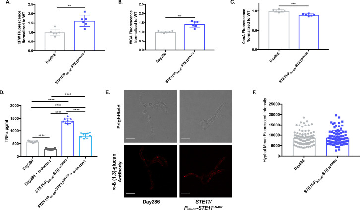
Masking the immunogenic cell wall epitope ß(1,3)-glucan under an outer layer of mannosylated glycoproteins is an important virulence factor deployed by Candida albicans during infection. Consequently, increased ß(1,3)-glucan exposure (unmasking) reveals C. albicans to the host's immune system and attenuates its virulence. We have previously shown that activation of the Cek1 MAPK pathway via expression of a hyperactive allele of an upstream kinase (STE11ΔN467) induced unmasking. It also increased survival of mice in a murine disseminated candidiasis model and attenuated kidney fungal burden by ≥33 fold. In this communication, we utilized cyclophosphamide-induced immunosuppression to test if the clearance of the unmasked STE11ΔN467 mutant was dependent on the host immune system. Suppression of the immune response by cyclophosphamide reduced the attenuation in fungal burden caused by the STE11ΔN467 allele. Moreover, specific depletion of neutrophils via 1A8 antibody treatment also reduced STE11ΔN467-dependent fungal burden attenuation, but to a lesser extent than cyclophosphamide, demonstrating an important role for neutrophils in mediating fungal clearance of unmasked STE11ΔN467 cells. In an effort to understand the mechanism by which Ste11ΔN467 causes unmasking, transcriptomics were used to reveal that several components in the Cek1 MAPK pathway were upregulated, including the transcription factor CPH1 and the cell wall sensor DFI1. In this report we show that a cph1ΔΔ mutation restored ß(1,3)-glucan exposure to wild-type levels in the STE11ΔN467 strain, confirming that Cph1 is the transcription factor mediating Ste11ΔN467-induced unmasking. Furthermore, Cph1 is shown to induce a positive feedback loop that increases Cek1 activation. In addition, full unmasking by STE11ΔN467 is dependent on the upstream cell wall sensor DFI1. However, while deletion of DFI1 significantly reduced Ste11ΔN467-induced unmasking, it did not impact activation of the downstream kinase Cek1. Thus, it appears that once stimulated by Ste11ΔN467, Dfi1 activates a parallel signaling pathway that is involved in Ste11ΔN467-induced unmasking.


Conflict of interest statement

The authors have declared that no competing interests exist.

Figures

Fig 1
Fig 1. Systemic infection with a hyperactive STE11ΔN467 mutant attenuates fungal burden in multiple niches within a mouse host.
(A) ICR mice were intravenously infected with 1x10⁶ cells of C. albicans wild-type (Day286) or the STE11/Ptet-off-STE11ΔN467 strain and their kidneys, livers, spleens, and brains were harvested 4 days post infection (d.p.i.) to assess fungal burden. (n = 6 mice)(*p<0.05, **p<0.01, ***p<0.0005, ****p<0.0001, by Kruskal-Wallis test with Dunn’s multiple comparisons post-hoc analysis). (B) Serum collected 4 days post infection was assessed to determine the protein concentrations of TNFα, CCL5, CXCL10, IFNα and IL-6 via flow cytometry using the LEGENDplex cytokine bead-based array kit. (n = 8 mice)(*p<0.05, **p<0.005, ***p<0.0005, by Mann-Whitney test).
Fig 2
Fig 2. Virulence attenuation during systemic infection by the hyperactive STE11ΔN467 mutant is host immune system dependent.
(A) ICR mice were immunosuppressed with recurring intraperitoneal injections of 150mg/kg of cyclophosphamide (Cyclo) every 3 days starting 4 days prior to infection. At day 0, mice were intravenously injected with 1x10⁴ cells of C. albicans wild-type (Day286) or the STE11/Ptet-off-STE11ΔN467 strain that were previously grown in YPD containing doxycycline to repress STE11ΔN467 expression prior to infection. Kidneys were harvested 4 days post infection (d.p.i.) to assess fungal burden. (B) Kidney fungal burden in mice immunosuppressed with cyclophosphamide 4 d.p.i. (n = 8–9 mice) (*p<0.0001, by student’s t-test). (C) Survival curve for cyclophosphamide-immunosuppressed mice that were intravenously injected with 1x10⁴ cells of wild-type (Day286) or the STE11/Ptet-off-STE11ΔN467 strain. Immunosuppression was maintained via recurring intraperitoneal injections every 3 days with 150mg/kg of cyclophosphamide starting 4 days prior to infection (day -4). (n = 10 mice) (D) Neutrophil depletion was achieved with recurring intraperitoneal injections of 300ug of anti-mouse Ly6G (1A8) antibody every 2 days starting one day prior to infection. At day 0, ICR mice were intravenously injected with 1x10⁴ cells of C. albicans wild-type (Day286) or the STE11/Ptet-offf-STE11ΔN467 strain and kidney fungal burden was assessed 4 d.p.i. (E) Kidney fungal burden in neutrophil depleted mice 4 d.p.i. (+/- Doxy = addition or absence of doxycycline) (n = 5 mice for Day286 and STE11/Ptet-off-STE11ΔN467 + doxycycline controls, n = 9 mice for STE11/Ptet-off-STE11ΔN467 –doxycycline, and n = 10 mice for Day286 –doxycycline) (***p<0.0005 and ****p<0.0001, by Kruskal-Wallis test with Dunn’s multiple comparisons post-hoc analysis).
Fig 3
Fig 3. Hyperactive STE11ΔN467 expression induces changes in cell wall components of yeast, but not hyphal cells of C. albicans.
(A-C) Overnight cultures of Day286 wild-type and STE11/Ptet-off-STE11ΔN467 cells grown in YPD without doxycycline were stained with calcofluor white (CFW), fluorescein conjugated wheat germ agglutinin (WGA) and Alexa Fluor 647 conjugated concanavalin A (ConA) to assess total chitin, surface exposed chitin, and mannan, respectively. Three biological replicates with 2 technical replicates were run for each sample. (A) CFW staining, (B) WGA staining and (C) ConA staining. (**p<0.005, ***p<0.0005, by student’s t-test). (D) The impact that dectin-1 recognition of exposed ß(1,3)-glucan has on macrophage activation was assessed via antibody blocking using an anti-dectin-1 monoclonal neutralizing antibody. RAW264.7 macrophages and UV-inactivated Day286 wild-type or STE11/Ptet-off-STE11ΔN467 cells were co-incubated for 4 hours at a multiplicity of infection (M.O.I.) of 10. Prior to infection, macrophages were either pretreated with 5ug/ml of the anti-dectin-1 neutralizing antibody or a corresponding volume of tissue culture media lacking the antibody. Following co-incubation with yeast cells, supernatants were collected and filtered through a 0.22μm filter and secreted TNFα levels were determined via an ELISA. (****p<0.0001, by one-way ANOVA with Tukey’s post hoc analysis). (E-F) Hyphal cells of the Day286 wild-type and the STE11/Ptet-off-STE11ΔN467 strain were stained with anti-ß(1,3)-glucan antibody and a Cy3-conjugated secondary antibody for microscopic analysis of hyphal ß(1,3)-glucan unmasking. (E) Representative confocal microscopy image of stained hyphal cells. The scale bar indicates 10μm. (F) Quantification of hyphal immunofluorescence via ImageJ analysis (n = 3 independent replicates for each strain with 30 hyphal cells quantified per replicate)(p = 0.9258, by student’s t-test).
Fig 4
Fig 4. Disruption of downstream components in the Cek1 MAPK pathway blocks ß(1,3)-glucan unmasking by hyperactive Ste11ΔN467.
(A-B) Overnight cultures of cells were stained with anti-ß(1,3)-glucan antibody and a phycoerythrin-conjugated secondary antibody followed by flow cytometry analysis to assess the levels of ß(1,3)-glucan exposure. (A) ß(1,3)-glucan unmasking of cek1ΔΔ mutants. (B) ß(1,3)-glucan unmasking of cph1ΔΔ mutants. (C-E) Overnight cultures of cells were stained with calcofluor white (CFW), fluorescein conjugated wheat germ agglutinin (WGA) and Alexa Fluor 647 conjugated concanavalin A (ConA) to assess total chitin, surface exposed chitin, and mannan levels, respectively, of cph1ΔΔ mutants. (C) CFW staining, (D) WGA staining and (E) ConA staining. (F) Overnight cultures were stained to assess ß(1,3)-glucan unmasking of the PENO1-CPH1 overexpression mutant. For all stains, three biological replicates with 2–3 technical replicates were analyzed by flow cytometry for each sample. (**p<0.01, ***p<0.001, ****p<0.0001, by one-way ANOVA with Tukey’s post hoc analysis).
Fig 5
Fig 5. CPH1 deletion reduces Ste11ΔN467 induced TNFα secretion by macrophages and partially restores virulence during systemic infection in mice.
(A) TNFα secretion by RAW264.7 macrophages. RAW264.7 murine macrophages and UV-inactivated C. albicans strains were co-incubated for 4 hours at a multiplicity of infection (M.O.I.) of 10. Culture supernatant was subsequently filtered through a 0.22μm filter and assessed via ELISA analysis to determine secreted TNFα levels. (*p = 0.0063, ****p<0.0001, by one-way ANOVA with Tukey’s post hoc analysis)(n = 3 biological replicates with 3 technical replicates for each sample). (B) Kidney fungal burden 4 d.p.i. during infection with cph1ΔΔ mutants. ICR mice were intravenously injected with 1x10⁶ cells of C. albicans wild-type (SC5314), STE11/Ptet-off-STE11ΔN467, STE11/Ptet-off-STE11ΔN467cph1ΔΔ or the STE11/Ptet-off-STE11ΔN467cph1ΔΔ::CPH1-Flag strain and kidneys were harvested 4 d.p.i to assess fungal burden. (n = 7–8 mice)(*p<0.05, ***p<0.001, ****p<0.0001, by Kruskal-Wallis test with Dunn’s multiple comparisons post-hoc analysis.).
Fig 6
Fig 6. Cph1 mediates activation of its upstream MAPK, Cek1.
Proteins were harvested from cells grown to mid-log phase for western blot analysis. Membranes were blotted using an anti-P44/42 antibody for phosphorylated Cek1 detection and an anti-Cek1 antibody for total Cek1 detection. (A) Western blot showing active and total Cek1 levels in cph1ΔΔ deletion mutants. (B) Fold change relative to the wild-type in Phosphorylated-Cek1 to total Cek1 levels for all CPH1 deletion mutants. (n = 3 biological replicates assessed on 3 separate western blots) (****p<0.0001, by one-way ANOVA with Tukey’s post hoc analysis). (C) Western blot showing active and total Cek1 levels for the PENO1-CPH1 overexpression mutant. (D) Fold change relative to the wild-type in Phosphorylated-Cek1 to total Cek1 levels for the PENO1-CPH1 overexpression mutant. (n = 3 biological replicates assessed on 3 separate western blots) (****p<0.0001, by students t-test).
Fig 7
Fig 7. DFI1 expression levels partially mediate ß(1,3)-glucan unmasking and macrophage TNFα secretion induced by hyperactive STE11ΔN467 expression.
(A-D) Overnight cultures of cells were stained with anti-ß(1,3)-glucan antibody and a phycoerythrin-conjugated secondary antibody for flow cytometry analysis or a Cy3-conjugated secondary antibody for confocal microscopy to assess the levels of ß(1,3)-glucan exposure. (A) ß(1,3)-glucan unmasking of opy2ΔΔ mutants. (B) ß(1,3)-glucan unmasking of dfi1ΔΔ mutants. (C) ß(1,3)-glucan unmasking of PENO1-DFI1 overexpression mutants. (****p<0.0001, by one-way ANOVA with Tukey’s post hoc analysis)(n = 3 biological replicates with 2–3 technical replicates were run for each sample.). (D) Representative microscopy images of ß(1,3)-glucan exposure of DFI1 overexpression mutants. The scale bar indicates 10μm. (E-F) The impact of altered DFI1 expression levels on macrophage TNFα secretion was assessed via co-incubation of RAW264.7 murine macrophages with UV-inactivated DFI1 mutants for 4 hours at a multiplicity of infection (M.O.I.) of 10. Culture supernatant was subsequently filtered through a 0.22um filter and assessed via ELISA analysis to determine secreted TNFα levels. (E) TNFα levels during co-incubation with RAW264.7 and dfi1ΔΔ mutants. (F) TNFα levels during co-incubation with RAW264.7 and PENO1-DFI1 overexpression mutants. (***p<0.0005, ****p<0.0001, by one-way ANOVA with Tukey’s post hoc analysis) (n = 3 biological replicates with 3 technical replicates for each sample).
Fig 8
Fig 8. Dfi1 does not mediate Ste11ΔN467-induced activation of the MAPK Cek1.
Proteins were harvested from cells grown to mid-log phase for western blot analysis. Membranes were blotted using an anti-P44/42 antibody for phosphorylated Cek1 detection and an anti-Cek1 antibody for total Cek1 detection. (A) Western blot showing active and total Cek1 levels in DFI1 deletion mutants. (B) Western blot showing active and total Cek1 levels in DFI1 overexpression mutants.
Fig 9
Fig 9. Proposed model for hyperactive STE11ΔN467 induced unmasking.
In this model, expression of the hyperactive STE11ΔN467 allele (red box) induces activation of the canonical Cek1 MAPK pathway (Ste11-Hst7-Cek1) and activates the downstream transcription factor Cph1. Cph1 activation in turn induces increased ß(1,3)-glucan exposure through regulated expression of genes within its regulon, and mediates a positive feedback loop into the pathway to further hyperactivate the upstream MAPK Cek1 and increase its transcriptional output. Simultaneously, Cph1 activation causes an upregulation in the transcript levels of the cell wall sensor DFI1, which is then stimulated as a result of hyperactivation of the Cek1 MAPK pathway and initiates signaling in a parallel, unidentified, pathway that is also necessary to achieve the full levels of unmasking during STE11ΔN467 expression.

References

    1. Brown GD, Denning DW, Gow NA, Levitz SM, Netea MG, White TC. Hidden killers: human fungal infections. Sci Transl Med. 2012;4(165):165rv13. Epub 2012/12/21. doi: 10.1126/scitranslmed.3004404. - DOI - PubMed
    1. Netea MG, Joosten LA, van der Meer JW, Kullberg BJ, van de Veerdonk FL. Immune defence against Candida fungal infections. Nat Rev Immunol. 2015;15(10):630–42. Epub 2015/09/22. doi: 10.1038/nri3897 . - DOI - PubMed
    1. Gow NAR, Latge JP, Munro CA. The Fungal Cell Wall: Structure, Biosynthesis, and Function. Microbiol Spectr. 2017;5(3). Epub 2017/05/18. doi: 10.1128/microbiolspec.FUNK-0035-2016. - DOI - PMC - PubMed
    1. Hopke A, Brown AJP, Hall RA, Wheeler RT. Dynamic Fungal Cell Wall Architecture in Stress Adaptation and Immune Evasion. Trends Microbiol. 2018;26(4):284–95. Epub 2018/02/18. doi: 10.1016/j.tim.2018.01.007 ; PubMed Central PMCID: PMC5869159. - DOI - PMC - PubMed
    1. Wheeler RT, Kombe D, Agarwala SD, Fink GR. Dynamic, morphotype-specific Candida albicans beta-glucan exposure during infection and drug treatment. PLoS Pathog. 2008;4(12):e1000227. Epub 2008/12/06. doi: 10.1371/journal.ppat.1000227; PubMed Central PMCID: PMC2587227. - DOI - PMC - PubMed

Publication types

Supplementary concepts