

Uterine leiomyosarcoma: a rare clinical entity
- PMID: 34433536
- PMCID: PMC8388282
- DOI: 10.1136/bcr-2021-244233
Uterine leiomyosarcoma: a rare clinical entity
Abstract
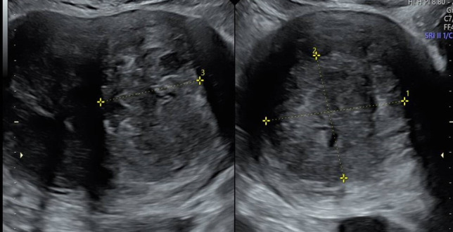
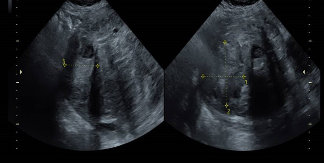
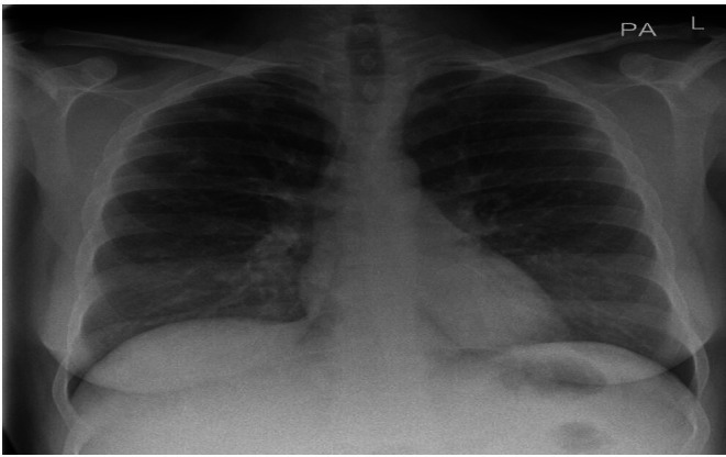
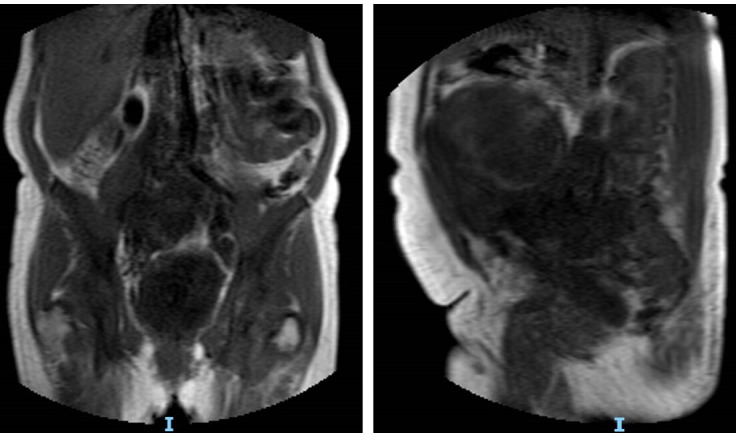
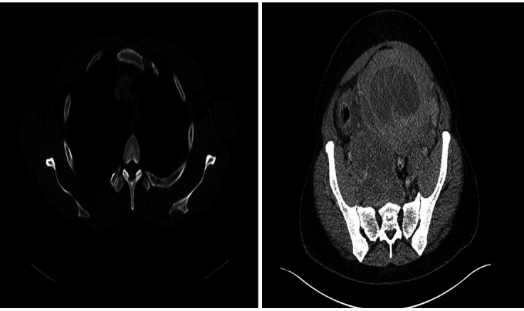
Leiomyosarcoma is a rare aggressive malignant mesenchymal tumour, accounting for 1% of all uterine malignancies. It spreads rapidly through the intraperitoneal and haematogenous pathways. It is often diagnosed postoperatively following myomectomy, hysterectomy or supracervical hysterectomy for presumed benign disease. It has a predilection for perimenopausal women with a median age of 50 years. Individuals may describe symptoms of vaginal or abdominal pressure. Physical examination may reveal a large palpable pelvic mass, which may haemorrhage. Surgery remains the mainstay of treatment. Hysterectomy and a bilateral salpingo-oophorectomy may be considered, depending on the individual's menopausal status. Ovarian preservation can be considered in young patients. Adjuvant systemic treatment and radiotherapy are of no benefit. Gemcitabine/docetaxel and doxorubicin have shown benefit in the treatment of advanced or recurrent disease. The authors present the case of a 44-year-old woman with lower abdominal pain, vaginal bleeding and a uterine fibroid. Laboratory investigations confirmed a leucocytosis, neutrophilia and a thrombocythaemia. Further investigation with an MRI pelvis showed a very large, heterogeneous, malignant appearing pelvic mass compressing the right ureter and it appeared uterine in nature. Her staging CT showed multiple lung metastases. The diagnosis of uterine leiomyosarcoma was subsequently established. Due to the aggressive behaviour of this sarcoma subtype, novel early detection strategies and targeted therapies are required.
Keywords: cancer - see oncology; gynecological cancer.
© BMJ Publishing Group Limited 2021. No commercial re-use. See rights and permissions. Published by BMJ.
Conflict of interest statement
Competing interests: None declared.
Figures
References
-
- Oliva E, Carcangiu ML, Carinelli SG, et al. . Mesenchymal tumours [chapter 5: tumours of the uterine corpus]. In: WHO classification of tumours of female reproductive organs. 4th edn. Lyon: IARC, 2014: 135–47.
Publication types
MeSH terms
LinkOut - more resources
Full Text Sources
Medical