Case Reports
. 2021 Nov;20(11):2098-2109.
doi: 10.1158/1535-7163.MCT-21-0361. Epub 2021 Aug 25.

Vimseltinib: A Precision CSF1R Therapy for Tenosynovial Giant Cell Tumors and Diseases Promoted by Macrophages

Affiliations
Case Reports

Vimseltinib: A Precision CSF1R Therapy for Tenosynovial Giant Cell Tumors and Diseases Promoted by Macrophages

Bryan D Smith et al. Mol Cancer Ther. 2021 Nov.

Abstract

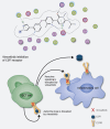
Macrophages can be co-opted to contribute to neoplastic, neurologic, and inflammatory diseases. Colony-stimulating factor 1 receptor (CSF1R)-dependent macrophages and other inflammatory cells can suppress the adaptive immune system in cancer and contribute to angiogenesis, tumor growth, and metastasis. CSF1R-expressing osteoclasts mediate bone degradation in osteolytic cancers and cancers that metastasize to bone. In the rare disease tenosynovial giant cell tumor (TGCT), aberrant CSF1 expression and production driven by a gene translocation leads to the recruitment and growth of tumors formed by CSF1R-dependent inflammatory cells. Small molecules and antibodies targeting the CSF1/CSF1R axis have shown promise in the treatment of TGCT and cancer, with pexidartinib recently receiving FDA approval for treatment of TGCT. Many small-molecule kinase inhibitors of CSF1R also inhibit the closely related kinases KIT, PDGFRA, PDGFRB, and FLT3, thus CSF1R suppression may be limited by off-target activity and associated adverse events. Vimseltinib (DCC-3014) is an oral, switch control tyrosine kinase inhibitor specifically designed to selectively and potently inhibit CSF1R by exploiting unique features of the switch control region that regulates kinase conformational activation. In preclinical studies, vimseltinib durably suppressed CSF1R activity in vitro and in vivo, depleted macrophages and other CSF1R-dependent cells, and resulted in inhibition of tumor growth and bone degradation in mouse cancer models. Translationally, in a phase I clinical study, vimseltinib treatment led to modulation of biomarkers of CSF1R inhibition and reduction in tumor burden in TGCT patients.


Figures

None
Graphical abstract
Figure 1. Structure of vimseltinib. A, Chemical structure of vimseltinib. B, Cocrystal structure of vimseltinib bound to CSF1R. Direct inhibitor hydrogen bonds with CSF1R. C, Nucleation of hydrogen bond network stabilized by vimseltinib binding. PDB:7MFC, (resolution 2.8 Å).
Figure 1.
Structure of vimseltinib. A, Chemical structure of vimseltinib. B, Cocrystal structure of vimseltinib bound to CSF1R. Direct inhibitor hydrogen bonds with CSF1R. C, Nucleation of hydrogen bond network stabilized by vimseltinib binding. PDB:7MFC, (resolution 2.8 Å).
Figure 2. Cellular activity of vimseltinib. A, Time course of inhibition of CSF1R phosphorylation in THP1 cells after compound washout. Cells were treated with 1 µmol/L vimseltinib for 2 hours prior to washout. CSF1R phosphorylation was detected by ELISA. B, Inhibition of M-NFS-60 cell proliferation. C, Effect of CSF1 ligand concentration on inhibition of M-NFS-60 cell proliferation by vimseltinib and pexidartinib. D, Inhibition of CSF1-stimulated phosphorylation of ERK downstream of CSF1R signaling by vimseltinib and pexidartinib in human whole blood.
Figure 2.
Cellular activity of vimseltinib. A, Time course of inhibition of CSF1R phosphorylation in THP1 cells after compound washout. Cells were treated with 1 µmol/L vimseltinib for 2 hours prior to washout. CSF1R phosphorylation was detected by ELISA. B, Inhibition of M-NFS-60 cell proliferation. C, Effect of CSF1 ligand concentration on inhibition of M-NFS-60 cell proliferation by vimseltinib and pexidartinib. D, Inhibition of CSF1-stimulated phosphorylation of ERK downstream of CSF1R signaling by vimseltinib and pexidartinib in human whole blood.
Figure 3. Pharmacokinetics/pharmacodynamics of vimseltinib in a mouse model. A, Steady-state inhibition of CSF1-stimulated cFOS expression after 6 days of oral dosing. B, Pharmacokinetic parameters of vimseltinib at steady state in mice. AUC(0–24) = area under the plasma concentration–time curve from 0–24 hours; Cl_obs = total body clearance; Cmax = maximum concentration; Tmax = time to maximum concentration; T½ = half-life; Vz_obs = volume of distribution. C, Plot of individual mouse spleen cFOS expression versus vimseltinib plasma concentration, with EC50 = 430 ng/mL.
Figure 3.
Pharmacokinetics/pharmacodynamics of vimseltinib in a mouse model. A, Steady-state inhibition of CSF1-stimulated cFOS expression after 6 days of oral dosing. B, Pharmacokinetic parameters of vimseltinib at steady state in mice. AUC(0–24) = area under the plasma concentration–time curve from 0–24 hours; Cl_obs = total body clearance; Cmax = maximum concentration; Tmax = time to maximum concentration; T½ = half-life; Vz_obs = volume of distribution. C, Plot of individual mouse spleen cFOS expression versus vimseltinib plasma concentration, with EC50 = 430 ng/mL.
Figure 4. Vimseltinib inhibition of bone degradation. A, Representative images are shown of the right hind limbs from 2 vehicle-treated mice and 2 mice treated with vimseltinib at 10 mg/kg every day in the PC3 peritibial bone invasion mouse model. B, Qualitative analysis of CT images of right hind limbs of all animals on study (n = 20/cohort).
Figure 4.
Vimseltinib inhibition of bone degradation. A, Representative images are shown of the right hind limbs from 2 vehicle-treated mice and 2 mice treated with vimseltinib at 10 mg/kg every day in the PC3 peritibial bone invasion mouse model. B, Qualitative analysis of CT images of right hind limbs of all animals on study (n = 20/cohort).
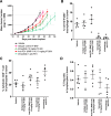
Figure 5. Vimseltinib inhibition of tumor growth and effects on the immune system in the syngeneic MC38 colorectal cancer mouse model. A, Mean tumor burden of treatment groups (n = 10/cohort). Mice were dosed days 12–39. B, Levels of macrophages in excised tumors after 3 weeks of treatment. C, Levels of CD8+ cytotoxic T cells in excised tumors after 3 weeks of treatment. D, Levels of Tregs in excised tumors after 3 weeks of treatment.
Figure 5.
Vimseltinib inhibition of tumor growth and effects on the immune system in the syngeneic MC38 colorectal cancer mouse model. A, Mean tumor burden of treatment groups (n = 10/cohort). Mice were dosed days 12–39. B, Levels of macrophages in excised tumors after 3 weeks of treatment. C, Levels of CD8+ cytotoxic T cells in excised tumors after 3 weeks of treatment. D, Levels of Tregs in excised tumors after 3 weeks of treatment.
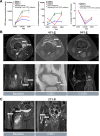
Figure 6. Preliminary biomarker and clinical activity of vimseltinib in patients with TGCT. A, Increases in the CSF1R ligands CSF1 and IL34 in plasma, and decrease of levels of CSF1R-dependent nonclassical monocytes in blood in vimseltinib-treated patients. Decrease in tumor burden in patients with TGCT, in the right knee of patient 1 (B) and the right hip of patient 2 (C). Patient 3 did not consent to release of MRI images.
Figure 6.
Preliminary biomarker and clinical activity of vimseltinib in patients with TGCT. A, Increases in the CSF1R ligands CSF1 and IL34 in plasma, and decrease of levels of CSF1R-dependent nonclassical monocytes in blood in vimseltinib-treated patients. Decrease in tumor burden in patients with TGCT, in the right knee of patient 1 (B) and the right hip of patient 2 (C). Patient 3 did not consent to release of MRI images.

Comment in

  • p. 2095

References

    1. Wynn TA, Chawla A, Pollard JW. Macrophage biology in development, homeostasis and disease.Nature 2013;496:445–55. - PMC - PubMed
    1. DeNardo DG, Brennan DJ, Rexhepaj E, Ruffell B, Shiao SL, Madden SF, et al. . Leukocyte complexity predicts breast cancer survival and functionally regulates response to chemotherapy.Cancer Discov 2011;1:54–67. - PMC - PubMed
    1. Mitchem JB, Brennan DJ, Knolhoff BL, Belt BA, Zhu Y, Sanford DE, et al. . Targeting tumor-infiltrating macrophages decreases tumor-initiating cells, relieves immunosuppression, and improves chemotherapeutic responses.Cancer Res 2013;73:1128–41. - PMC - PubMed
    1. Wyckoff JB, Wang Y, Lin EY, Li JF, Goswami S, Stanley ER, et al. . Direct visualization of macrophage-assisted tumor cell intravasation in mammary tumors.Cancer Res 2007;67:2649–56. - PubMed
    1. Ruffell B, Coussens LM. Macrophages and therapeutic resistance in cancer.Cancer Cell 2015;27:462–72. - PMC - PubMed

Publication types

Substances