. 2021 Aug 31;118(35):e2101287118.
doi: 10.1073/pnas.2101287118.

An in vitro vesicle formation assay reveals cargo clients and factors that mediate vesicular trafficking

Affiliations

An in vitro vesicle formation assay reveals cargo clients and factors that mediate vesicular trafficking

Yan Huang et al. Proc Natl Acad Sci U S A. .

Abstract

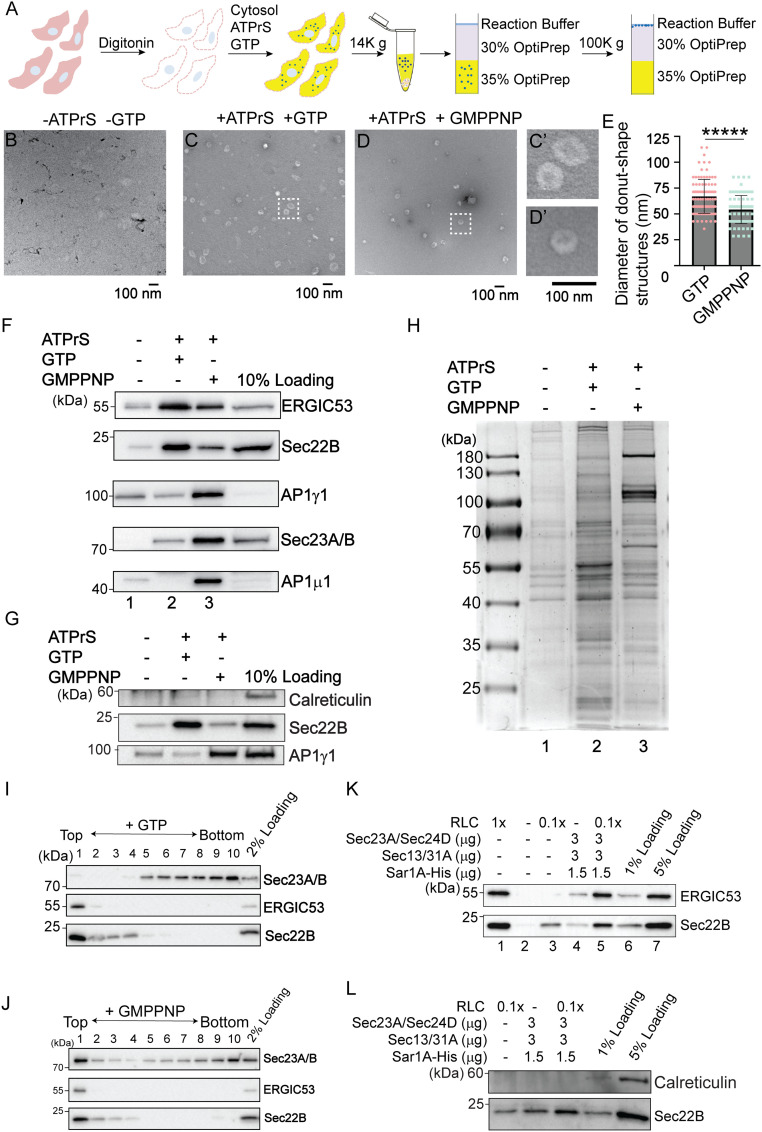
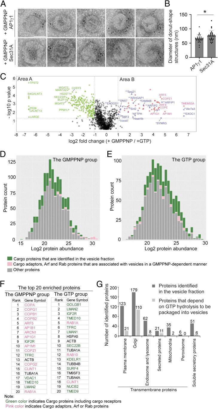
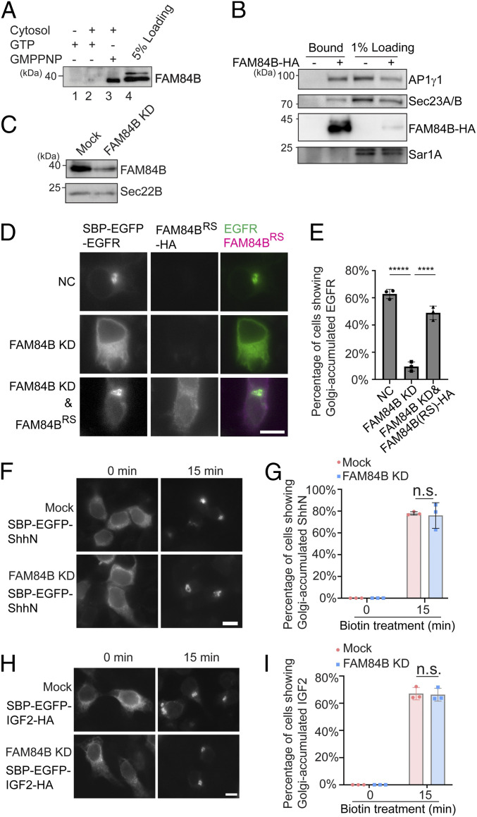
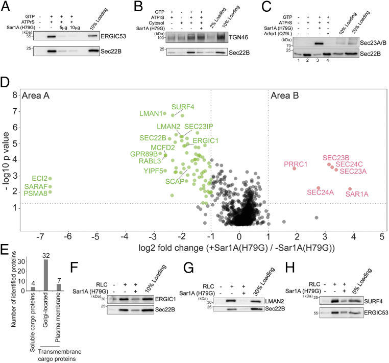
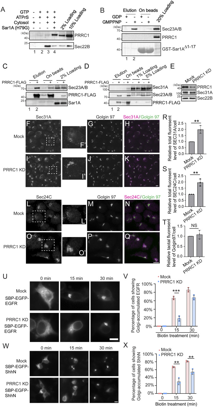
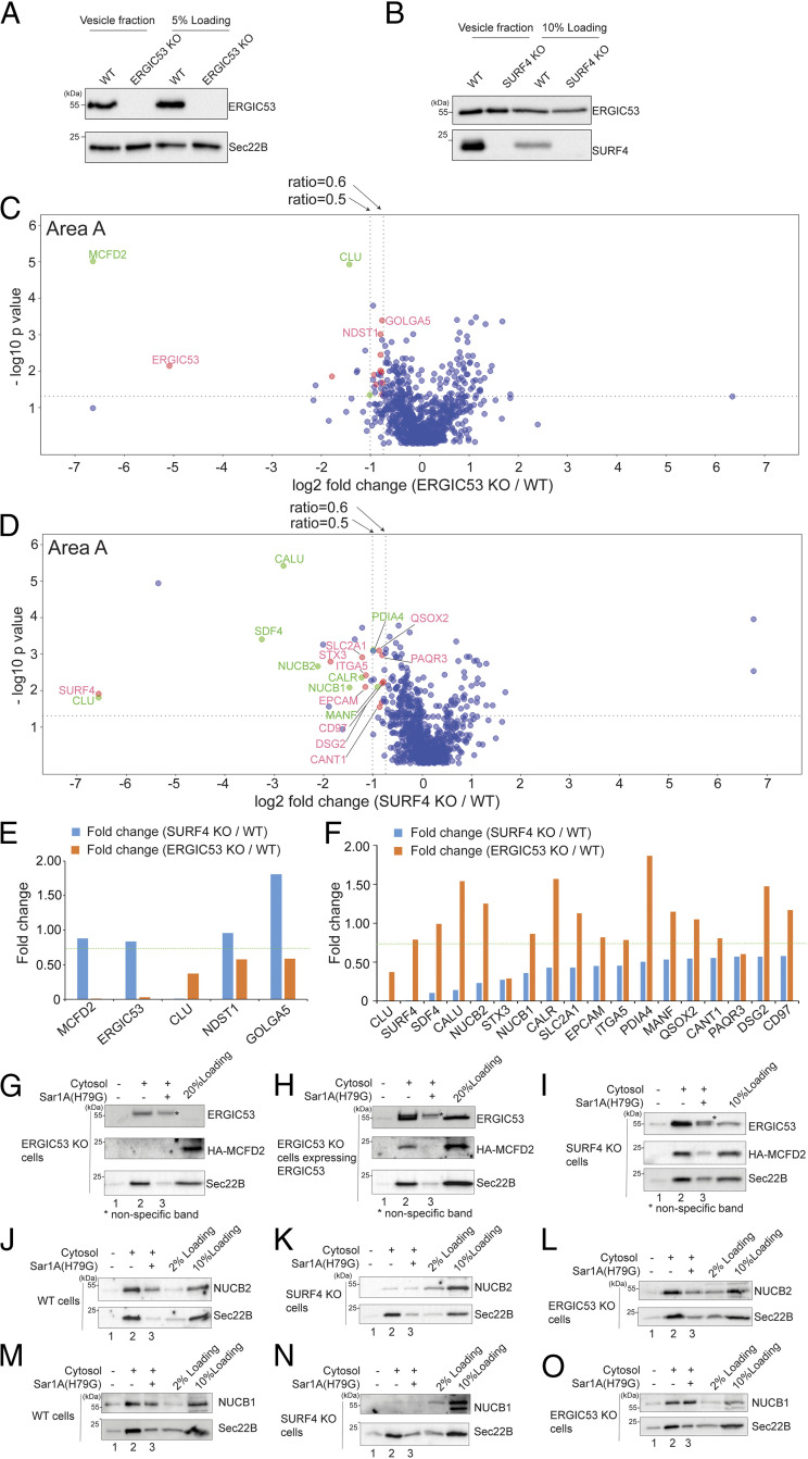
The fidelity of protein transport in the secretory pathway relies on the accurate sorting of proteins to their correct destinations. To deepen our understanding of the underlying molecular mechanisms, it is important to develop a robust approach to systematically reveal cargo proteins that depend on specific sorting machinery to be enriched into transport vesicles. Here, we used an in vitro assay that reconstitutes packaging of human cargo proteins into vesicles to quantify cargo capture. Quantitative mass spectrometry (MS) analyses of the isolated vesicles revealed cytosolic proteins that are associated with vesicle membranes in a GTP-dependent manner. We found that two of them, FAM84B (also known as LRAT domain containing 2 or LRATD2) and PRRC1, contain proline-rich domains and regulate anterograde trafficking. Further analyses revealed that PRRC1 is recruited to endoplasmic reticulum (ER) exit sites, interacts with the inner COPII coat, and its absence increases membrane association of COPII. In addition, we uncovered cargo proteins that depend on GTP hydrolysis to be captured into vesicles. Comparing control cells with cells depleted of the cargo receptors, SURF4 or ERGIC53, we revealed specific clients of each of these two export adaptors. Our results indicate that the vesicle formation assay in combination with quantitative MS analysis is a robust and powerful tool to uncover novel factors that mediate vesicular trafficking and to uncover cargo clients of specific cellular factors.

Keywords: COPII; cargo receptor; cargo sorting; intracellular protein transport; secretory pathway.


Conflict of interest statement

The authors declare no competing interest.

Figures

Fig. 1.
Fig. 1.
A large-scale in vitro vesicle formation assay for proteomic analysis. (A) Diagram demonstrating the experimental procedures for the vesicle formation assay. (B–D) Visualization of the morphology of the buoyant membrane structures formed in the budding reaction. The buoyant membranes were isolated by density gradient flotation and analyzed by negative staining TEM. (C′ and D′) The magnified views of the indicted areas in C and D. (Scale bar, 100 nm.) (E) Quantification of the diameter of donut shape structures from three biological repeats (mean ± SD *****P < 0.00001). (F–H) The vesicle formation assay was performed using the indicated reagents. Vesicle fractions were analyzed by immunoblot (F and G) or Coomassie blue staining (H). ATPrS: ATP regeneration system. (I and J) The vesicle formation assay was performed in the presence of GTP (I) or GMPPNP (J). The vesicle fractions were evaluated by density gradient flotation. (K and L) The vesicle formation assay was performed using the indicated reagents. The vesicle fraction was analyzed by immunoblot using the indicated antibodies. Data shown in F, G, and K are representative example of three biological repeats.
Fig. 2.
Fig. 2.
Identification of cytosolic proteins that are associated with vesicles in a GTP-dependent manner and cargo proteins that are packaged into vesicles in a GTP-hydrolysis–dependent manner. (A) Immunogold TEM was performed using AP1γ1 and Sec31A antibodies to label the donut shape structures produced in the presence of GMPPNP. (Scale bar, 50 nm.) (B) Quantification of the diameter of the donut shape structures labeled by antibodies against AP1γ1 and Sec31A (mean ± SD *P < 0.05, from two biological repeats). (C) The vesicle formation assay was performed in the presence of GTP or GMPPNP. The isolated vesicles in each experimental group were resuspended in RapiGest SF surfactant. The proteins in the vesicle fractions were trypsin digested and analyzed by label-free mass spectrometry. A total of 1,285 proteins were identified in both experimental groups. The log2 ratio of the abundance of each identified protein in the vesicles prepared in the presence of GMPPNP over that in the vesicles prepared in the presence of GTP was plotted on the x axis and the –log10 P value of the difference was plotted on the y axis. (D and E) Histogram of the log2 abundance of the human proteins identified in the vesicle fraction produced in the presence of GMPPNP (D) or GTP (E). (F) The list of the top 20 abundant human proteins in the GMPPNP group or in the GTP group. (G) Number of proteins categorized based on predictions from Uniprot.
Fig. 3.
Fig. 3.
FAM84B/LRATD2 is recruited to vesicle membranes in a GTP-dependent manner, interacts with AP1γ1 and Sec23A/B, and regulates ER-to-Golgi transport of EGFR but not ShhN or IGF2. (A) The vesicle formation assay was performed using the indicated reagents. The proteins in the vesicle fraction were analyzed by Western blot. (B) HEK293T cells expressing the FAM84B-HA were treated in 2 mM dithiobis(succinimidyl propionate) (DSP), and the cell lysates were incubated with beads conjugated with HA antibodies. After incubation, the bound proteins were analyzed by Western blot using the indicated antibodies. (C) HEK293T cells were transfected with control siRNA or siRNA against FAM84B/LRATD2. Day 3 after transfection, cells were lysed and analyzed by Western blot. (D, F, and H) HEK293T cells were transfected with control siRNA or siRNA against FAM84B/LRATD2. Twenty-four hours after transfection, cells were transfected with plasmids encoding the indicted constructs. On day 3 after knockdown, cells were incubated with biotin and cycloheximide for 15 min and the localization of the cargo proteins was analyzed by fluorescent microscope. (Scale bar, 10 μm.) (E, G, and I) Quantifications of the percentage of cells showing Golgi-localized cargo proteins in each experimental group (mean ± SD; n = 3; >100 cells counted for each experiment). ****P < 0.0001; *****P < 0.00001; n.s., not significant. Data shown in A–C are representative examples of three biological repeats.
Fig. 4.
Fig. 4.
Identification of cargo proteins and cytosolic proteins that are dependent on Sar1A to be associated with transport vesicles. (A–C) The vesicle formation assay was performed using the indicated reagents. Vesicle fractions were analyzed by immunoblot. (D) The vesicle formation assay was performed in the presence or absence of Sar1A(H79G). The isolated vesicles in each experimental group were resuspended in RapiGest SF surfactant. The proteins in the vesicle fractions were trypsin digested and analyzed by label-free mass spectrometry. The log2 ratio of the abundance of each identified protein in the vesicles prepared in the presence of Sar1A(H79G) over that in the vesicles prepared in the absence of Sar1A(H79G) was plotted on the x axis and the −log10 P value of the difference was plotted on the y axis. (E) Number of proteins identified in area A in D categorized based on predictions from Uniprot. (F–H) The vesicle formation assay was performed using the indicated reagents. The vesicle fraction was analyzed by immunoblot. Data shown in A–C and F–H are representative examples of three biological repeats.
Fig. 5.
Fig. 5.
PRRC1 interacts with Sec23A/B and knockdown increases membrane association of Sec31A and Sec24C and decreases ER-to-Golgi transport of EGFR and ShhN. (A) The vesicle formation assay was performed using the indicated reagents. Vesicle fractions were analyzed by immunoblot. (B) GST-Sar1AΔ1–17 was loaded with GDP or GMPPNP and then incubated with rat liver cytosol. After incubation, proteins that bound to Sar1A in a nucleotide-dependent manner were eluted with EDTA. The eluted fraction and the proteins left on beads after elution were analyzed by immunoblot. (C and D) M2 agarose beads were incubated with cell lysates from HEK293T cells expressing the PRRC1-FLAG. After incubation, the bound proteins were eluted with 3× FLAG peptides and analyzed by Western blot using the indicated antibodies. (E) HEK293T cells were transfected with control siRNA or siRNA against PRRC1. Day 3 after transfection, cells were lysed and analyzed by Western blot. (FQ) HEK293T cells were transfected with control siRNA (F–H, LN) or siRNA against PRRC1 (I–K, OQ). Day 3 after transfection, the localizations of Sec31A, Sec24C, and Golgin97 were analyzed by immunofluorescence. (Scale bar, 10 μm.) The magnified views of the indicated area in F, I, L, and O are shown in F′, I′, L′, and O′. (R–T) Quantifications of the total fluorescent level of Sec31A (R), Sec24C (S), and Golgin97 (T) per cell (mean ± SD; n = 3; >125 cells from nine random imaging fields counted for each experiment). In each experiment, the total fluorescent level was normalized to that in mock cells. **P < 0.01; NS, not significant. (U and W) HEK293T cells were transfected with control siRNA or siRNA against PRRC1. Twenty-four hours after transfection, cells were retransfected with plasmids encoding the indicated construct. On day 3 after knockdown, cells were incubated with biotin for the indicated time and the localization of the indicated protein was analyzed by fluorescent microscope. (Scale bar, 10 μm.) (V and X) Quantifications of the percentage of cells showing Golgi-localized EGFR or ShhN in cells treated with control siRNA or siRNA against PRRC1 (mean ± SD; n = 3; >100 cells counted for each experiment). ***P < 0.001; **P < 0.01. Data shown in A, B, D, and E are representative examples of three biological repeats.
Fig. 6.
Fig. 6.
Identification of cargo proteins that depend on ERGIC53 or SURF4 for being packaged into transport vesicles. (A and B) The vesicle formation was performed using wild-type HEK293TRex cells and ERGIC53 KO HEK293TRex cells (A) or SURF4 KO HEK293TRex cells (B). The vesicle fraction was then analyzed by immunoblot. (C and D) The vesicle formation assay was performed using wild-type HEK293TRex cells and ERGIC53 KO HEK293TRex cells (C) or SURF4 KO HEK293TRex cells (D). The isolated vesicles in each experimental group were resuspended in RapiGest SF surfactant. The proteins in the vesicle fractions were trypsin digested and analyzed by label-free mass spectrometry. The log2 ratio of the abundance of each identified protein in the vesicles prepared from ERGIC53 KO or SURF4 KO cells over that in the vesicles prepared in wild-type cells was plotted on the x axis and the −log10 P value of the difference was plotted on the y axis. (E) The fold change of the identified ERGIC53 client was compared with the fold change of the abundance of these proteins in the vesicle fraction prepared from the WT cells and SURF4 KO cells. (F) The fold change of identified SURF4 client was compared with the fold change of the abundance of these proteins in the vesicle fraction prepared from the WT cells and ERGIC53 KO cells. Dotted line indicates the fold change of 0.75. (G–O) The vesicle formation was performed using the indicated cells. Vesicle fractions were then analyzed by immunoblot. Data shown in GO are representative examples of three biological repeats. The single asterisks in panels GI indicate the nonspecific bands recognized by anti-ERGIC53 antibodies.

References

    1. Guo Y., Sirkis D. W., Schekman R., Protein sorting at the trans-Golgi network. Annu. Rev. Cell Dev. Biol. 30, 169–206 (2014). - PubMed
    1. Lee M. C., Miller E. A., Goldberg J., Orci L., Schekman R., Bi-directional protein transport between the ER and Golgi. Annu. Rev. Cell Dev. Biol. 20, 87–123 (2004). - PubMed
    1. Dancourt J., Barlowe C., Protein sorting receptors in the early secretory pathway. Annu. Rev. Biochem. 79, 777–802 (2010). - PubMed
    1. Nichols W. C., et al. ., Mutations in the ER-Golgi intermediate compartment protein ERGIC-53 cause combined deficiency of coagulation factors V and VIII. Cell 93, 61–70 (1998). - PubMed
    1. Vollenweider F., Kappeler F., Itin C., Hauri H. P., Mistargeting of the lectin ERGIC-53 to the endoplasmic reticulum of HeLa cells impairs the secretion of a lysosomal enzyme. J. Cell Biol. 142, 377–389 (1998). - PMC - PubMed

Publication types