. 2021 Sep 20;97(12):e1170-e1181.
doi: 10.1212/WNL.0000000000012580.

Acute and Chronic Effects of Multiple Concussions on Midline Brain Structures

Affiliations

Acute and Chronic Effects of Multiple Concussions on Midline Brain Structures

Nathan W Churchill et al. Neurology. .

Abstract

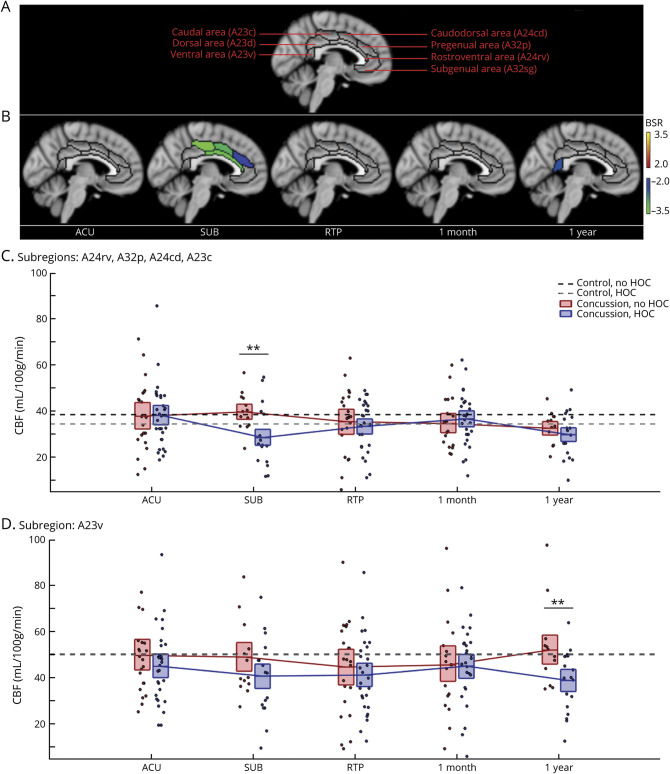
Background and objectives: To test the hypothesis that a history of concussion (HOC) causes greater disturbances in cerebral blood flow (CBF) and white matter microstructure of midline brain structures after subsequent concussions, during the acute and chronic phases of recovery.

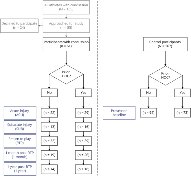
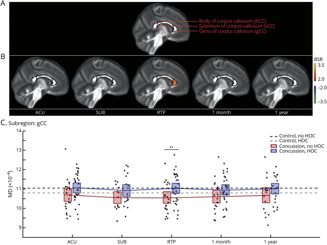
Methods: In this longitudinal MRI study, 61 athletes with uncomplicated concussion (36 with HOC) were imaged at the acute phase of injury (1-7 days after injury), the subacute phase (8-14 days), medical clearance to return to play (RTP), 1 month after RTP, and 1 year after RTP. A normative group of 167 controls (73 with HOC) were also imaged. Each session assessed CBF of the cingulate cortex, along with fractional anisotropy (FA) and mean diffusivity (MD) of the corpus callosum. Linear mixed models tested for interactions of HOC with time since injury. The Sport Concussion Assessment Tool (SCAT) was also used to evaluate effects of HOC on symptoms, cognition, and balance.

Results: Athletes with HOC had significantly greater declines in midcingulate CBF subacutely (z = -3.29, p = 0.002) and greater declines in posterior cingulate CBF at 1 year after RTP (z = -2.42, p = 0.007). No significant effects of HOC were seen for FA, whereas athletes with HOC had higher MD of the splenium at RTP (z = 2.54, p = 0.008). These effects were seen in the absence of significant differences in SCAT domains (|z| ≤ 1.14, p ≥ 0.256) or time to RTP (z = 0.23, p = 0.818).

Discussion: Results indicate subacute and chronic effects of HOC on cingulate CBF and callosal microstructure in the absence of differences in clinical indices. These findings provide new insights into physiologic brain recovery after concussion, with cumulative effects of repeated injury detected among young, healthy athletes.


Figures

Figure 1
Figure 1. Study Flowchart Reporting Sample Sizes at Each Imaging Session
HOC = history of concussion.
Figure 2
Figure 2. Effects of HOC on Post-concussion Cerebral Blood Flow
(A) Segmentation of the cingulate cortex based on the Brainnetome Atlas into 7 subregions. (B) For cingulate regions and imaging sessions showing significant history of concussion (HOC) effects on cerebral blood flow (CBF) during concussion recovery, standardized effects are shown as bootstrap ratios (BSRs). (C) Distribution of concussed athlete CBF values averaged over subregions showing significant effects at subacute injury (SUB), as denoted by double asterisks. (D) Distribution of concussed athlete CBF values for the subregion showing significant effects at 1 year after return to play (RTP), as denoted by double asterisks. For distribution plots, concussed athletes without and with HOC are plotted separately. Horizontal red/blue lines denote group means, and boxes indicate 95% confidence intervals of the mean. Distribution means are connected between sessions by solid red/blue lines, and the mean CBF values for controls without and with HOC are plotted as black and gray horizontal dashed lines, respectively. ACU = acute phase of injury.
Figure 3
Figure 3. Effects of HOC on post-concussion mean diffusivity (MD)
(A) Segmentation of the corpus callosum based on the John Hopkins University atlas into 3 subregions. (B) For callosal regions and imaging sessions showing significant history of concussion (HOC) effects on mean diffusivity (MD) during concussion recovery, standardized effects are shown as bootstrap ratios (BSRs). (C) Distribution of concussed athlete MD values for the subregion showing effects at return to play (RTP), as denoted by double asterisks. For distribution plots, concussed athletes without and with HOC are plotted separately. Horizontal red/blue lines denote group means, and boxes indicate 95% confidence intervals. Distribution means are connected between sessions by solid red/blue lines, and the mean MD values for controls without and with HOC are plotted as black and gray horizontal dashed lines, respectively. ACU = acute phase of injury; SUB = subacute injury.

Comment in

References

    1. Churchill NW, Hutchison MG, Graham SJ, Schweizer TA. Mapping brain recovery after concussion: from acute injury to one year after medical clearance. Neurology. 2019;93(21):e1980-e1992. - PMC - PubMed
    1. Churchill NW, Hutchison MG, Richards D, Leung G, Graham SJ, Schweizer TA. Neuroimaging of sport concussion: persistent alterations in brain structure and function at medical clearance. Sci Rep. 2017;7(1):8297. - PMC - PubMed
    1. Guskiewicz KM, Marshall SW, Bailes J, et al. . Association between recurrent concussion and late-life cognitive impairment in retired professional football players. Neurosurgery. 2005;57(4):719-726. - PubMed
    1. Guskiewicz KM, Marshall SW, Bailes J, et al. . Recurrent concussion and risk of depression in retired professional football players. Med Sci Sports Exerc. 2007;39(6):903-909. - PubMed
    1. Gavett BE, Stern RA, Cantu RC, Nowinski CJ, McKee AC. Mild traumatic brain injury: a risk factor for neurodegeneration. Alzheimers Res Ther. 2010;2(3):18. - PMC - PubMed