Observational Study
. 2021 Aug 25;12(1):5119.
doi: 10.1038/s41467-021-25441-6.

Portable, bedside, low-field magnetic resonance imaging for evaluation of intracerebral hemorrhage

Affiliations
Observational Study

Portable, bedside, low-field magnetic resonance imaging for evaluation of intracerebral hemorrhage

Mercy H Mazurek et al. Nat Commun. .

Abstract

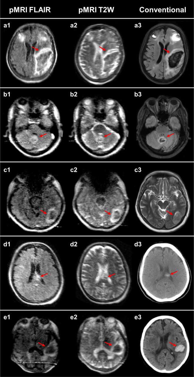
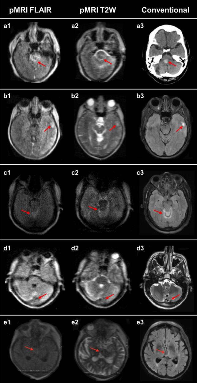
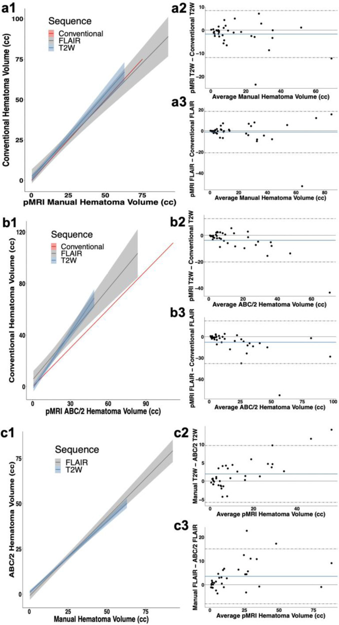
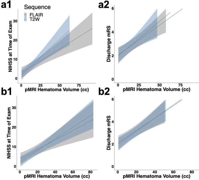
Radiological examination of the brain is a critical determinant of stroke care pathways. Accessible neuroimaging is essential to detect the presence of intracerebral hemorrhage (ICH). Conventional magnetic resonance imaging (MRI) operates at high magnetic field strength (1.5-3 T), which requires an access-controlled environment, rendering MRI often inaccessible. We demonstrate the use of a low-field MRI (0.064 T) for ICH evaluation. Patients were imaged using conventional neuroimaging (non-contrast computerized tomography (CT) or 1.5/3 T MRI) and portable MRI (pMRI) at Yale New Haven Hospital from July 2018 to November 2020. Two board-certified neuroradiologists evaluated a total of 144 pMRI examinations (56 ICH, 48 acute ischemic stroke, 40 healthy controls) and one ICH imaging core lab researcher reviewed the cases of disagreement. Raters correctly detected ICH in 45 of 56 cases (80.4% sensitivity, 95%CI: [0.68-0.90]). Blood-negative cases were correctly identified in 85 of 88 cases (96.6% specificity, 95%CI: [0.90-0.99]). Manually segmented hematoma volumes and ABC/2 estimated volumes on pMRI correlate with conventional imaging volumes (ICC = 0.955, p = 1.69e-30 and ICC = 0.875, p = 1.66e-8, respectively). Hematoma volumes measured on pMRI correlate with NIH stroke scale (NIHSS) and clinical outcome (mRS) at discharge for manual and ABC/2 volumes. Low-field pMRI may be useful in bringing advanced MRI technology to resource-limited settings.


Conflict of interest statement

K.N.S. is the principal investigator. This study received support from the Collaborative Science Award from the American Heart Association (PIs: K.N.S., W.T.K., M.S.R.), National Institutes of Health Supplement Grant, and Hyperfine Research, Inc. research grant. W.T.K. receives grants from NIH and AHA; grants and personal fees from Biogen, Inc; grants and personal fees from NControl Therapeutics; has a patent pending that is licensed to NControl Therapeutics; holds equity in Woolsey Pharmaceuticals. M.S.R. is a co-founder of Hyperfine Research, Inc. J.R. is a co-founder of Hyperfine Research, Inc. E.B.W., L.S., and M.P. are research scientists and engineers at Hyperfine Research, Inc. All other authors declare no competing interests.

Figures

Fig. 1
Fig. 1. Portable (0.064T) magnetic resonance imaging device dimensions.
a The portable MRI (pMRI) device has a height of 140 cm and a width of 86 cm. The critical 5 Gauss (0.5 mT) boundary around the scanner extends into a circle with a diameter of 158 cm. b The pMRI device is positioned at the head of the patient’s hospital bed. The scanner bridge (35 cm) adjoins the hospital bed with the pMRI device and the patient’s chest height and head and neck lengths are positioned within the vertical clearance between magnets (32 cm) and the head coil length (26 cm), respectively. c The patient’s head is positioned within the single channel transmit, 8-channel receiver head coil (26 × 20 cm) and the RF shield is closed for scan acquisition, which creates a horizontal clearance of 55 cm.
Fig. 2
Fig. 2. Intracerebral hemorrhage at 0.064T versus conventional imaging modalities (CT or 3T MRI).
The first and second columns are low-field FLAIR and T2W images, respectively. The third column is a gold-standard clinical examination for comparison (3T MRI: a3, b3, c3, and e3; CT: d3). a Left isointense fronto-parietal intracerebral hemorrhage (ICH) with hyperintense rim and bilateral frontal hematomas. b Bilateral isointense cerebellar ICH with hyperintense rim. c Left hypointense occipital lobe ICH with hyperintense rim. d Left homogenous, hyperintense ICH in corpus collosum. e Left hypointense temporal ICH with hyperintense rim.
Fig. 3
Fig. 3. False negative intracerebral hemorrhage cases.
The first and second columns are low-field FLAIR and T2W images, respectively. The third column is a gold-standard clinical examination for comparison. (3T MRI: b3, d3, and e3; CT: a3). a Right cerebellar pontine intracerebral hemorrhage (ICH). Missed by all raters. b Left temporal ICH. Missed by all raters. c Bilateral cerebellar ICH. Missed by 2/3 raters. d Left cerebellum ICH. Missed by all raters. e Left thalamus ICH. Missed by all raters.
Fig. 4
Fig. 4. Hematoma volume measurements on portable MRI.
a1 Validation of manually segmented pMRI hematoma volumes against manual volumes on conventional (CT or 1.5/3T MRI) imaging (T2W (n = 37): r = 0.952, 95% CI: [0.907–0.975], p < 2.20e-16; FLAIR (n = 38): r = 0.899, 95% CI: [0.812–0.946], p = 1.90e-14). Bland-Altman plots for manual pMRI showed a bias of −1.70 cc [limits of agreement (LOA): −11.8–8.42] for (a2) T2W sequences (n = 37) and a bias of −1.22 cc [LOA: −20.8–18.4] for (a3) FLAIR (n = 38). b1 Validation of averaged ABC/2 estimated pMRI volumes against averaged estimated volumes on conventional (CT or 1.5/3T MRI) imaging (T2W (n = 40): r = 0.945, 95% CI: [0.892–0.972], p < 2.20e-16; FLAIR (n = 38): r = 0.835, 95% CI: [0.702–0.911], p = 7.53e-11). Bland–Altman plots for ABC/2 pMRI showed a bias of −3.74 cc [LOA: −20.2–12.7] for (b2) T2W (n = 40) and a bias of −7.89 [LOA: −38.0–22.2] for (b3) FLAIR (n = 38). c1 Manually segmented pMRI hematoma volumes against averaged estimated volumes using ABC/2 (T2W (n = 37): r = 0.977, 95% CI: [0.956–0.989], p < 2.20e-16; FLAIR (n = 38): r = 0.968, 95% CI: [0.936–0.984], p < 2.20e-16). Bland–Altman plots showed a bias of 1.962 [LOA: −5.76–9.68] for (c2) T2W (n = 37) and a bias of 1.79 [LOA: −4.46–8.04] for (c3) FLAIR (n = 38). Pearson correlations are reported for a1, b1, and c1 with confidence intervals. Line of identity shown in red (a1, b1). 95% confidence intervals are represented by bands (a1, b1, c1) and dashed gray lines (a2–3, b2–3, c2–3).
Fig. 5
Fig. 5. Hematoma volume and cognitive scores on portable MRI.
Manual pMRI hematoma volume versus (a1) cognitive status (NIHSS) at time of exam (pMRI T2W (n = 33): ρ = 0.750, 95% CI: [0.591–0.906], p = 4.95e-7; pMRI FLAIR (n = 34): ρ = 0.802, 95% CI: [0.669–0.930], p = 1.23e-8) and a2 functional status (mRS) at discharge (pMRI T2W (n = 36): ρ = 0.589, 95% CI: [0.372–0.804], p = 1.55e-4; pMRI FLAIR (n = 37): ρ = 0.641, 95% CI: [0.425–0.855], p = 1.89e-5). Averaged ABC/2 estimated pMRI hematoma volume versus (b1) cognitive status (NIHSS) at time of exam (pMRI T2W (n = 36): ρ = 0.805, 95% CI: [0.659–0.948], p = 3.18e-9; pMRI FLAIR (n = 34): ρ = 0.776, 95% CI: [0.617–0.930], p = 7.16e-8) and (b2) functional status (mRS) at discharge (pMRI T2W (n = 39): ρ = 0.747, 95% CI: [0.592–0.899], p = 4.85e-8; pMRI FLAIR (n = 37): ρ = 0.669, 95% CI: [0.483–0.853], p = 5.98e-6). Spearman correlations are reported for a1, a2, b1, and b2 with confidence intervals. Bands represent 95% confidence intervals.

References

    1. Kernan WN, et al. Guidelines for the prevention of stroke in patients with stroke and transient ischemic attack: a guideline for healthcare professionals from the American Heart Association/American Stroke Association. Stroke. 2014;45:2160–2236. doi: 10.1161/STR.0000000000000024. - DOI - PubMed
    1. Saver JL. Time is brain—quantified. Stroke. 2006;37:263–266. doi: 10.1161/01.STR.0000196957.55928.ab. - DOI - PubMed
    1. Campbell BC, Parsons MW. Imaging selection for acute stroke intervention. Int. J. Stroke. 2018;08:554–567. doi: 10.1177/1747493018765235. - DOI - PubMed
    1. Morgan CD, et al. Physiologic imaging in acute stroke: patient selection. Interv. Neuroradiol. 2015;21:499–510. doi: 10.1177/1591019915587227. - DOI - PMC - PubMed
    1. Powers WJ, et al. Guidelines for the early management of patients with acute ischemic stroke: 2019 update to the 2018 guidelines for the early management of acute ischemic stroke: A Guideline for Healthcare Professionals From the American Heart Association/American Stroke Association. Stroke. 2019;50:e344–e418. doi: 10.1161/STROKEAHA.118.022606. - DOI - PubMed

Publication types