Review
. 2021 Aug 9:12:716469.
doi: 10.3389/fimmu.2021.716469. eCollection 2021.

NF-κB: At the Borders of Autoimmunity and Inflammation

Affiliations
Review

NF-κB: At the Borders of Autoimmunity and Inflammation

Laura Barnabei et al. Front Immunol. .

Abstract

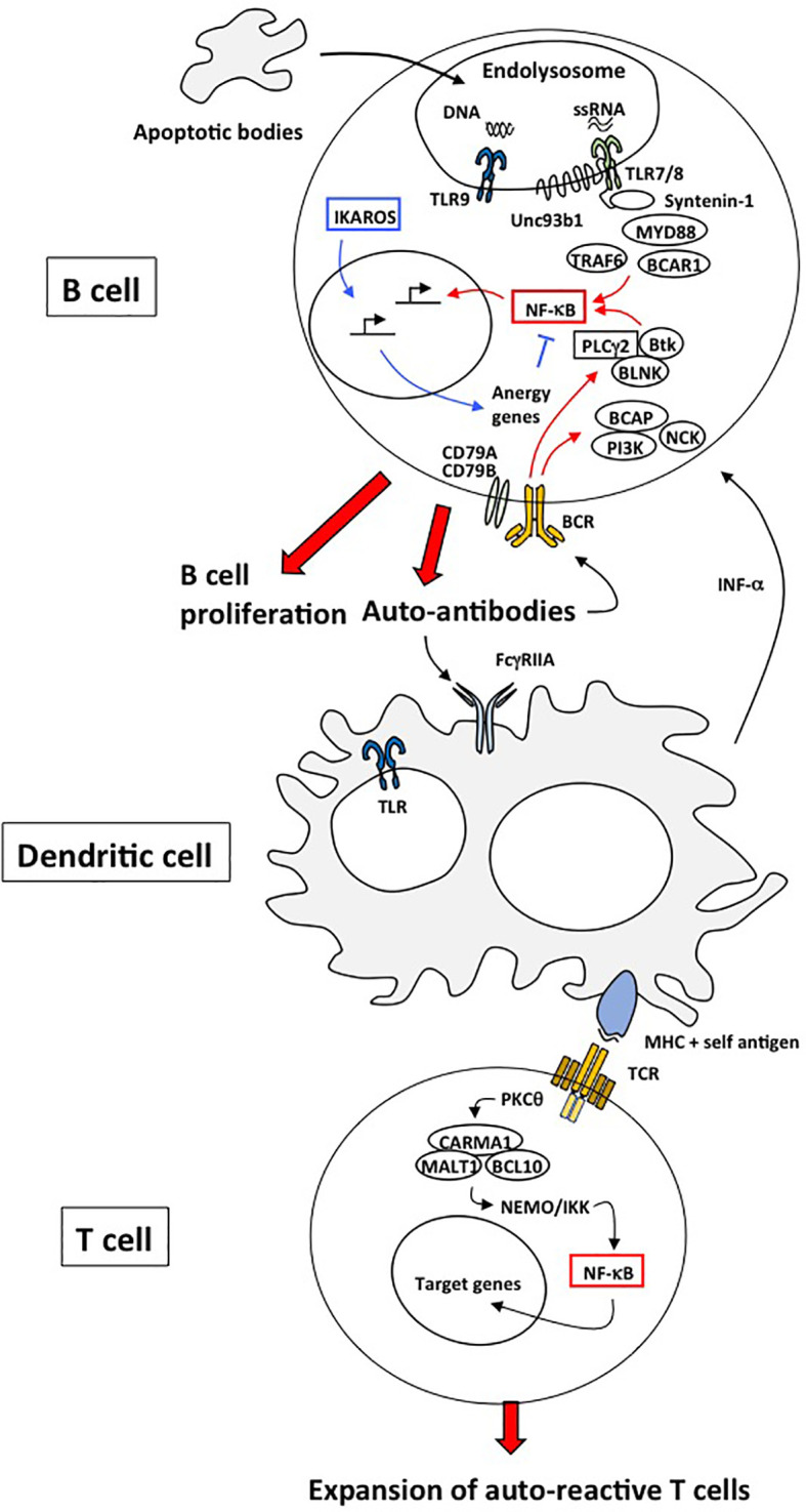
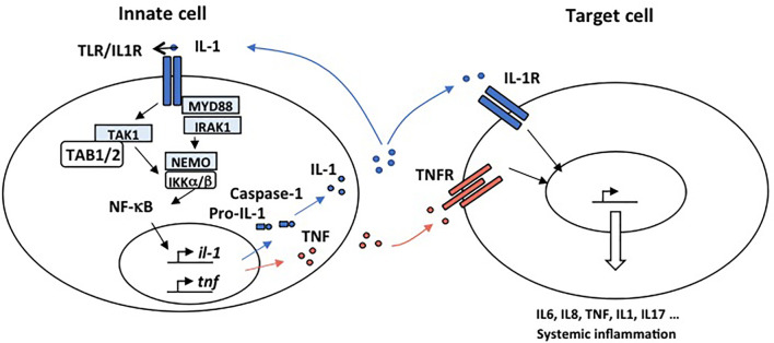
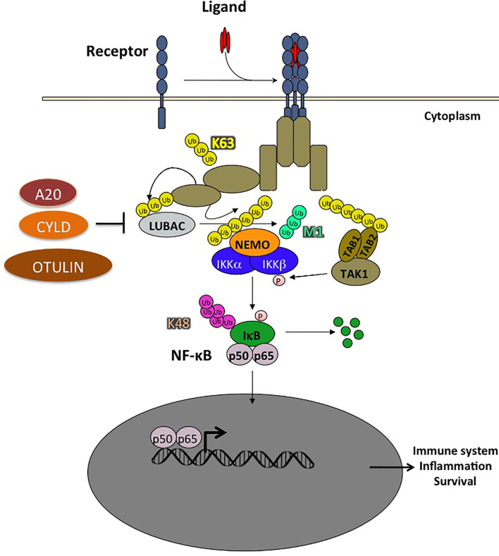
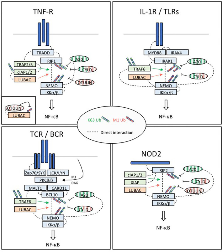
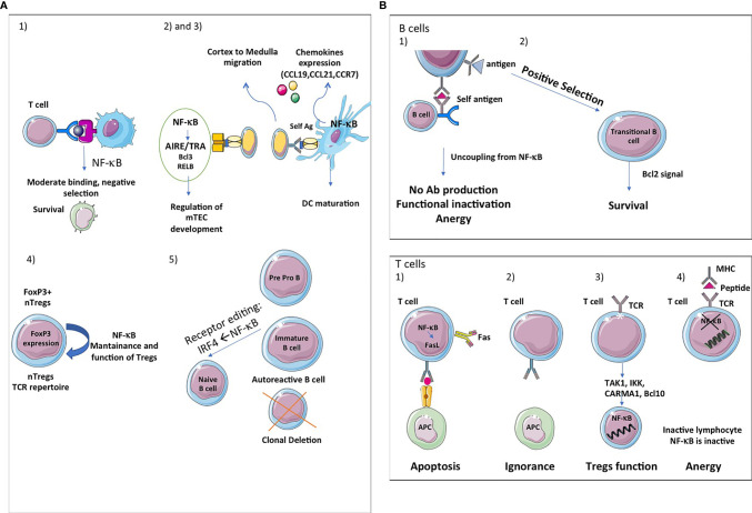
The transcription factor NF-κB regulates multiple aspects of innate and adaptive immune functions and serves as a pivotal mediator of inflammatory response. In the first part of this review, we discuss the NF-κB inducers, signaling pathways, and regulators involved in immune homeostasis as well as detail the importance of post-translational regulation by ubiquitination in NF-κB function. We also indicate the stages of central and peripheral tolerance where NF-κB plays a fundamental role. With respect to central tolerance, we detail how NF-κB regulates medullary thymic epithelial cell (mTEC) development, homeostasis, and function. Moreover, we elaborate on its role in the migration of double-positive (DP) thymocytes from the thymic cortex to the medulla. With respect to peripheral tolerance, we outline how NF-κB contributes to the inactivation and destruction of autoreactive T and B lymphocytes as well as the differentiation of CD4+-T cell subsets that are implicated in immune tolerance. In the latter half of the review, we describe the contribution of NF-κB to the pathogenesis of autoimmunity and autoinflammation. The recent discovery of mutations involving components of the pathway has both deepened our understanding of autoimmune disease and informed new therapeutic approaches to treat these illnesses.

Keywords: autoinflammation; genetic diseases; NF-κB; autoimmunity; immune tolerance; ubiquitination.


Conflict of interest statement

The authors declare that the research was conducted in the absence of any commercial or financial relationships that could be construed as a potential conflict of interest.

Figures

Figure 1
Figure 1
Mechanisms responsible for autoimmunity involving TLRs, BCR and TCR. In B cells, defective clearance of apoptotic cell debris releases nucleic acids capable of activating endosomal TLR7 and TLR9. TLR7- and TLR9-mediated NF-κB activation is dependent on MyD88, IRAK family of kinases and TRAF6. The chaperone UNC93B1 limits TLR7 signaling through its interaction with Syntenin-1 but not TLR9 and thus prevents TLR7-mediated autoimmunity. These TLR receptors can operate in conjunction with the BCR to activate NF-κB in order to increase the production of antibodies. Another transcription gene, Ikaros, induces the expression of anergy genes of which certain inhibit NF-κB. As a consequence, mutations of Ikaros or anergic genes can be responsible of autoimmune diseases. Dendritic cells induce peripheral tolerance by directing the fate of antigen-specific T cells. DCs can present self-antigens to T cells, providing transient T cell activation that can lead to either anergy or deletion of these T cells. DC-mediated tolerance is thus an active process that requires TCR-mediated NF-κB signaling.
Figure 2
Figure 2
Mechanisms responsible for autoimmunity involving IL-1R and TNFR. IL-1 and TNFα are proinflammatory cytokines able to promote systemic inflammation. Activation of the IL1R complex is part of the innate immune response and plays a major role in setting up the adaptive response. An “inflammatory cascade” leading to the cleavage of IL-1β by Caspase 1 activates this complex. They are important receptors for the initiation of inflammation. All IL-1R contain, like TLR, a TIR domain that intact with MyD88 through an homotypic interaction and drive NF-κB activation. IL-1 is well recognized for its role in the pathogenesis of disorders of autoinflammation. In autoinflammatory diseases, the effector cell is a myeloid cell, characteristically a monocyte or macrophage. IL-1 plays a key role in the expression of several pro-inflammatory cytokines. TNFα is a potent proinflammatory master cytokine modulating inflammatory processes, and its rapid induction is fundamental for the orchestration of the immune response. Monocytes and macrophages are important cellular mediators of TNF-mediated signal transduction. Chronic TNF stimulation leads to inflammatory diseases in which NF-κB is activated such as autoimmunity.
Figure 3
Figure 3
Ubiquitination plays a crucial role in NF-κB activation pathways. The ubiquitination of a substrate can take place with a single ubiquitin (monoubiquitination) or a chain of covalently linked polyubiquitin molecules (polyubiquitination). In the NF-κB pathway, the NF-κB inhibitor, IκB, is modified by K48-linked polyubiquitin chains which are recognized by the 26S proteasome, leading to the degradation of these proteins and the translocation of NF-κB into the nucleus. The polyubiquitin chains linked through the Lysine K63 of ubiquitin do not trigger the degradation of proteins, but rather have non-proteolytic functions such as protein trafficking or activation of kinases and phosphatases. In addition, ubiquitin chains can also be linked linearly, the C-terminal glycine being linked to an N-terminal methionine. Two RING type E3 ligases, HOIL1 and HOIP, specifically assemble linear polyubiquitin chains which play an important role in the regulation of NF-κB. Other types of ubiquitin modifications have also been observed but are not presented in this schema. Ubiquitination is reversible and counter-regulated by a family of deubiquitinases (DUB). There are 3 DUBs playing an important role in the negative regulation of the NF-κB pathway: A20, CYLD and OTULIN, the latter cleaving exclusively linear ubiquitin chains.
Figure 4
Figure 4
Regulation of NF-κB activation in four different pathways implicated in immune tolerance. TNFR: Upon binding of TNFα, the TNFR1 receptor trimerizes resulting in the recruitment of TRADD to the death domain (DD) of the receptors cytoplasmic tail. RIP1 is recruited through TRADD and TNFR1 through homotypic DD interactions. High affinity binding of TRAF2 trimers to TRADD is augmented by TRAF2/RIP1 interactions. TRAF2 trimers recruit cIAP1/2, which in turn recruit the LUBAC complex (HOIP, HOIL-1 and SHARPIN), while RIP1 mediates recruitment of TAK1 and the IKK complex through NEMO. This signaling complex (complex I) signals to NF-κB by recruiting in proximity TAK1 and the IKK complex supporting phosphorylation and activation of IKK. The ubiquitin ligase cIAP1/2 and LUBAC may facilitate TAK1 and IKK activation through production of linear and/or K63 linked ubiquitination. Then, activated IKK phosphorylates IκBα leading its ubiquitination and degradation and nuclear translocation of p65:p50 NF-κB complexes. DNA bound canonical NF-κB induces transcription of immune response genes as well as genes that protect the cell from TNF induced cell death. IL1R/TLRs: Despite differences in their extracellular domains, the IL-1 receptor (IL-1R) and TLRs contain a common cytoplasmic motif termed the Toll/IL-1R (TIR) homology domain, which is required for activation of NF-κB signaling pathways. IL-1R- and TLR-mediated NF-κB activation is initiated by the recruitment of MyD88 to the TIR. MyD88 is a scaffold protein that recruits the death domain-containing protein IRAK-1, IRAK-2, IRAK-4, and the ubiquitin protein ligase (E3) TRAF6. IRAK-4 and TRAF6 are essential signaling components of IL-1R- and TLR-mediated MAPK and NF-κB activation. IRAK-1 also plays an important role in IL-1R/TLR signaling in order to induce IKK activation and subsequently IκB activation and NF-κB nuclear translocation. TCR/BCR: In response to TCR or BCR triggering, phosphorylation of CARD11 (CARMA1) by PKC-θ or PKC-β on different Serine residues of its linker domain modifies its conformation allowing its association with a constitutively associated dimer formed by BCL10 and MALT1, leading to the assembly of the CBM complex. The formation of this complex constitutes one of the important steps towards NF-κB stimulation following TCR/BCR engagement. In turn, this complex activates the IκB kinase (IKK) responsible for the phosphorylation of the inhibitory factors IκBα and their subsequent degradation, allowing activation and nuclear translocation of NF-κB followed by transcription of its target genes. In addition to its scaffold function during NF-κB signaling, the paracaspase MALT1 exerts a proteolytic activity to ensure the regulation of NF-κB activation. NOD2: NOD2 immune function drive a higher incidence of autoimmune diseases such as Crohn’s disease. NOD2-mediated signaling relies on RIP2. RIP2 recruits a number of signaling regulators to the NOD2-associated protein complex, including several ubiquitin E3 ligases such as XIAP, c-IAP1/2, Pellino3, and LUBAC, which promote diverse ubiquitination of RIP2. XIAP is a critical ubiquitin ligase in the NOD2-RIP2 inflammatory pathway and promotes NEMO/IKK- mediated NF-κB activation.
Figure 5
Figure 5
NF-κB, a key player in central and peripheral immune tolerance. (A) Central tolerance: The thymic stroma is crucial for the growth, differentiation, positive and negative selection of the T-cell receptor repertoire. The thymic stromal compartment is composed of epithelium, fibroblasts, endothelium, macrophages and dendritic cells, each of which has a distinct role in T-cell development. Single-positive (SP) thymocytes after positive selection have to migrate into medulla to undergo negative selection and further maturation, given the critical role of mTECs in presenting tissue-restricted antigens (TRAs). The thymic medulla is indeed the main site for both negative selection of autoreactive thymocytes and positive selection of Tregs, two important central tolerance mechanisms. (1) First, the classical and alternative pathway of NF-κB activation contributes to the survival of immature cells during the negative selection and thereafter. (2) Second, the alternative NF-κB pathway regulates AIRE/TRAs expression and is involved in mTEC development, homeostasis and function. Bcl3 and RelB also regulate mTEC development at the DNA level. The deubiquitinase CylD is implicated in mTEC differentiation (3). Third, NF-κB also regulates the expression of chemokines (CCL19, CCL21, CCR7) implicated in cortex to medulla migration of DP thymocytes and to secondary lymphocytes organs and tissues. (4) Fourth, the NF-kB pathway has also been shown to influence thymic or natural Treg (nTreg) cells development and function. These cells recognize self-antigens and play an important role in the maintenance of immunological self-tolerance. Forkhead Box P3 (FOXP3) is the main transcription factor for the differentiation of these cells. NF-κB is important for the maintenance and functions of nTregs and the expression of FOXP3. (5) Fifth, in B cells, NF-κB participates in clonal deletion of autoreactive B lymphocytes and receptor editing, a protective mechanism of ongoing gene rearrangement that generates a new receptor with an innocuous specificity that prevents cell death by apoptosis. If the BCR recognizes sel-antigen, it is down regulated under the control of IRF4, a target gene of NF-κB. (B) Peripheral tolerance: Peripheral tolerance takes place in the periphery and its purpose is to ensure that T cells and B cells that escape from the thymus cannot give rise to autoimmunity. NF-κB signaling is essential for the development and maintenance of secondary and tertiary lymphoids organs. In B cells, peripheral tolerance uses two different mechanisms: (1) Anergy, which is characterized by desensitization of BCR signalling and its uncoupling from the NF-κB pathway, resulting in a failure of antigen presentation and antibody production. (2) NF-κB is essential for the survival of developing B cells in the spleen. Bcl2 is involved in the survival of transitional B cells. The classical and alternative pathways pathway of NF-κB activation are involved in B cells maturation. In T cells, peripheral tolerance uses four different mechanisms: (1) Inactivation of effector T cells by clonal deletion. Programmed cell death (apoptosis) is an essential mechanism leading to clonal deletion of T cells with high affinity for self-antigens. NF-κB plays a pro-apoptotic role in negative selection. (2) T-cells ignore certain self-antigens because they are located in immune-privileged sites or because they have low immunogenicity (low levels of expression or low binding affinity). (3) Conversion of T cells in Tregs. Non canonical NF-κB activation is important for the maintenance of these cells in vivo via its effect on mTEC development and canonical NF-κB plays is involved in Foxp3 expression which is fundamental for the function of Tregs. Components of the TCR-mediated NF-κB pathway such as TAK1, IKK, CARMA1 and Bcl10 are also involved in Tregs development. Ubiquitination is fundamental for Tregs functions. (4) Induction of anergy. Anergy is a tolerance mechanism in which lymphocytes are intrinsically functionally inactive. NF-κB is inactive in these unresponsive lymphocytes. NF-κB also controls DCs maturation.

References

    1. Liu T, Zhang L, Joo D, Sun SC. NF-kappaB Signaling in Inflammation. Signal Transduct Target Ther (2017) 2:17023–32. 10.1038/sigtrans.2017.23 - DOI - PMC - PubMed
    1. Oeckinghaus A, Ghosh S. The NF-kappaB Family of Transcription Factors and its Regulation. Cold Spring Harb Perspect Biol (2009) 1(4):a000034. 10.1101/cshperspect.a000034 - DOI - PMC - PubMed
    1. Savinova OV, Hoffmann A, Ghosh G. The Nfkb1 and Nfkb2 Proteins P105 and P100 Function as the Core of High-Molecular-Weight Heterogeneous Complexes. Mol Cell (2009) 34(5):591–602. 10.1016/j.molcel.2009.04.033 - DOI - PMC - PubMed
    1. Ghosh G, Wang VY, Huang DB, Fusco A. NF-kappaB Regulation: Lessons From Structures. Immunol Rev (2012) 246(1):36–58. 10.1111/j.1600-065X.2012.01097.x - DOI - PMC - PubMed
    1. Perkins ND. Integrating Cell-Signalling Pathways With NF-kappaB and IKK Function. Nat Rev Mol Cell Biol (2007) 8(1):49–62. 10.1038/nrm2083 - DOI - PubMed

Publication types

MeSH terms