Review
. 2021 Aug 19;9(8):23259671211021352.
doi: 10.1177/23259671211021352. eCollection 2021 Aug.

Current Concepts on Subtalar Instability

Affiliations
Review

Current Concepts on Subtalar Instability

Bruno S Pereira et al. Orthop J Sports Med. .

Abstract

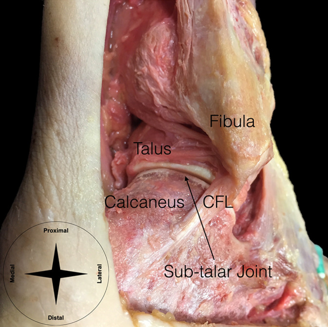
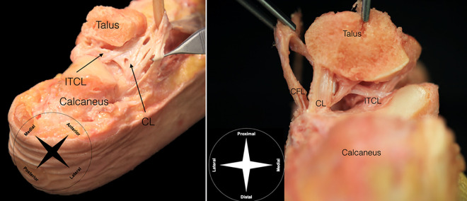
Subtalar instability remains a topic of debate, and its precise cause is still unknown. The mechanism of injury and clinical symptoms of ankle and subtalar instabilities largely overlap, resulting in many cases of isolated or combined subtalar instability that are often misdiagnosed. Neglecting the subtalar instability may lead to failure of conservative or surgical treatment and result in chronic ankle instability. Understanding the accurate anatomy and biomechanics of the subtalar joint, their interplay, and the contributions of the different subtalar soft tissue structures is fundamental to correctly diagnose and manage subtalar instability. An accurate diagnosis is crucial to correctly identify those patients with instability who may require conservative or surgical treatment. Many different nonsurgical and surgical approaches have been proposed to manage combined or isolated subtalar instability, and the clinician should be aware of available treatment options to make an informed decision. In this current concepts narrative review, we provide a comprehensive overview of the current knowledge on the anatomy, biomechanics, clinical and imaging diagnosis, nonsurgical and surgical treatment options, and outcomes after subtalar instability treatment.

Keywords: current concepts; instability; subtalar.


Conflict of interest statement

The authors declared that there are no conflicts of interest in the authorship and publication of this contribution. AOSSM checks author disclosures against the Open Payments Database (OPD). AOSSM has not conducted an independent investigation on the OPD and disclaims any liability or responsibility relating thereto.

Figures

Figure 1.
Figure 1.
Bony anatomy of the subtalar joint. CFL, calcaneofibular ligament.
Figure 2.
Figure 2.
Ligamentous anatomy of the subtalar joint. CFL, calcaneofibular ligament; CL, cervical ligament; ITCL, interosseous talocalcaneal ligament.
Figure 3.
Figure 3.
The anterolateral drawer test to assess subtalar joint laxity. The tibia is stabilized and the hindfoot is held with maximum ankle dorsiflexion to lock the joint in the ankle mortise and avoid tibiotalar movement. Then, (A) anterior stress is applied at the posterior calcaneus, (B) combined with inversion, internal rotation, and adduction of the forefoot, as shown by arrows.
Figure 4.
Figure 4.
The talar tilt test to assess subtalar joint laxity. The tibia and fibula are stabilized proximally and (A) a varus stress (arrow) is applied while holding the calcaneus for the talar tilt stress test or (B) it is applied a medially directed stress (arrow) while holding the calcaneus for the medial subtalar glide test.
Figure 5.
Figure 5.
Manual stress radiography under varus stress to assess talar tilt: (A) no stress and (B) with varus stress.
Figure 6.
Figure 6.
(A) T1-weighted MRI scan with a coronal view showing rupture of interosseous talocalcaneal ligament (arrow). (B) T2-weighted fat-suppressed MRI scan with a sagittal view showing absence of the anterior capsular ligament (arrow). MRI, magnetic resonance imaging.
Figure 7.
Figure 7.
(A) The nonanatomic Evans tenodesis procedure. The peroneus brevis muscle is separated, and its proximal end is sutured to the peroneus longus muscle. The peroneus brevis tendon is detached at the musculotendinous junction, but its distal insertion is preserved. A tunnel is created connecting the posterosuperior side of the fibula to the lateral malleolar tip, and the peroneus brevis tendon is routed through the tunnels from anterior to posterior and sutured on itself at the muscle belly. (B) The nonanatomic Chrisman-Snook procedure. Half of the peroneus brevis tendon is harvested proximally, but its distal attachment is preserved. A diagonal tunnel is made at the lateral malleolus, and another is made on the calcaneofibular ligament insertion at the calcaneus. The peroneus brevis graft is passed anteroposteriorly through the lateral malleolar tunnel, routed below the peroneal tendons, and then passed posteroanteriorly through the calcaneal tunnel. The graft is tensioned with the ankle in neutral flexion and rotation and sutured on itself. (C) The nonanatomic modified Elmslie procedure. Half of the peroneus brevis tendon is harvested proximally, but its distal attachment is preserved. A horizontal tunnel is made at the lateral malleolus, and another is made on the calcaneofibular ligament insertion at the calcaneus. The peroneus brevis graft is passed anteroposteriorly through the lateral malleolar tunnel, routed below the peroneal tendons, passed posteroanteriorly through the calcaneal tunnel, and then sutured on itself.
Figure 8.
Figure 8.
(A) Anatomic isolated reconstruction of the CL described by Schon et al. Half of the peroneus brevis tendon is harvested proximally, but its distal attachment is preserved. The graft is routed through the calcaneus vertical tunnel and passed through the talar neck tunnel, and then it is brought back and sutured on itself. (B) Triligamentous reconstruction using a plantaris tendon graft described by Schon et al. The graft is routed through a calcaneus (exiting at the posterior calcaneal tuberosity) and fibular tunnels, recreating the CFL and the ATFL, respectively. Then, it is passed through a tunnel at the talar neck (exiting at the floor of talar canal) and to a tunnel at the anterior and lateral calcaneus, replicating the CL. The graft is brought back by the same tunnels and secured to itself. (C) Triligamentous reconstruction, described by Schon et al, using half or the entire peroneus brevis tendon when the plantaris tendon is not available. The peroneus brevis tendon is harvested, but its distal insertion is preserved. The graft is routed through a tunnel at the anterior and lateral calcaneus (exiting at the floor of the talar canal) and then to a tunnel at the talar neck, replicating the CL. The graft is passed posteroinferiorly through a fibular tunnel, routed beneath the peroneal longus tendon then through a calcaneal tunnel (at the posterior calcaneal tuberosity), and attached to itself and the surrounding soft tissue. (D) ITCL anatomic reconstruction using a strip of the Achilles tendon described by Kato. The graft is passed though oblique tunnels at the calcaneus and the talar neck. The graft ends are fixed using staples. (E) ITCL anatomic reconstruction using half of the peroneus brevis tendon described by Pisani and Pisani et al. Half of the peroneus brevis is passed through the calcaneus and talar neck tunnels and then brought back through another calcaneal tunnel. The 2 tunnels aim to replicate the double-stranded ITCL. The graft end is looped and sutured on itself. ATFL, anterior talofibular ligament; CFL, calcaneofibular ligament; CL, cervical ligament; ITCL, interosseous talocalcaneal ligament.
Figure 9.
Figure 9.
Interosseous talocalcaneal ligament reconstruction using a suture-button fixation device. The tunnel placement can be safely achieved using a fluoroscopically guided percutaneous technique described by So et al. The graft is then fixed using a suture-button fixation device.
Figure 10.
Figure 10.
Anatomic reconstruction of subtalar ligaments using a semitendinosus allograft described by Jung et al. The allograft is passed through 2 fibular tunnels: one from the fibular distal tip to the posterior fibula (origin of the CFL) to the midfibular axis and another from above the origin of the ATFL, obliquely directed (45°) to the midfibular axis (1 cm proximal to the previous tunnel). The graft is then fixed using a 2-cm whip stitch at the posterior calcaneus and an interference screw at the anterior calcaneus. This technique aims to replicate the anterior and posterior subtalar ligaments in which the anterior limb assumes the function of the ITCL and the CL and the posterior limb anatomically reconstructs the CFL. ATFL, anterior talofibular ligament; CFL, calcaneofibular ligament; CL, cervical ligament; ITCL, interosseous talocalcaneal ligament.
Figure 11.
Figure 11.
Anatomic repair using the Broström-Gould procedure. The calcaneofibular ligament and the anterior talofibular ligament rupture ends are directly repaired and reinforced using the extensor retinaculum.
Figure 12.
Figure 12.
(A) The lateral view and (B) the anteroposterior view radiographs of a patient with subtalar dislocation.

References

    1. Agarwal S, Garg S, Vasudeva N. Subtalar joint instability and calcaneal spurs associated with the configuration of the articular facets of adult human calcaneum in Indian population. J Clin Diagn Res. 2016;10(9):AC05–AC09. - PMC - PubMed
    1. Al-Mohrej OA, Al-Kenani NS. Chronic ankle instability: current perspectives. Avicenna J Med. 2016;6(4):103–108. - PMC - PubMed
    1. Ashton-Miller JA, Ottaviani RA, Hutchinson C, Wojtys EM. What best protects the inverted weight-bearing ankle against further inversion? Evertor muscle strength compares favorably with shoe height, athletic tape, and 3 orthoses. Am J Sports Med. 1996;24(6):800–809. - PubMed
    1. Attarian DE, McCrackin HJ, DeVito DP, McElhaney JH, Garrett WE, Jr. Biomechanical characteristics of human ankle ligaments. Foot Ankle. 1985;6(2):54–58. - PubMed
    1. Aynardi M, Pedowitz DI, Raikin SM. Subtalar instability. Foot Ankle Clin. 2015;20(2):243–252. - PubMed