

Microscopic segmentation and classification of COVID-19 infection with ensemble convolutional neural network
- PMID: 34435702
- PMCID: PMC8646237
- DOI: 10.1002/jemt.23913
Microscopic segmentation and classification of COVID-19 infection with ensemble convolutional neural network
Abstract
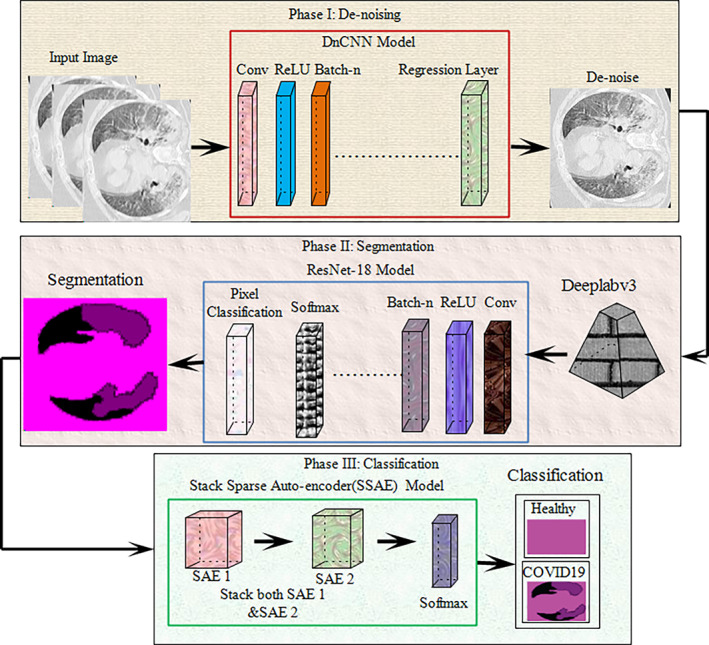
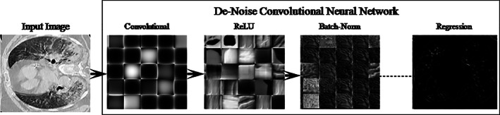
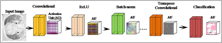
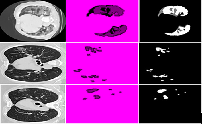
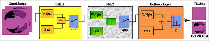
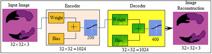
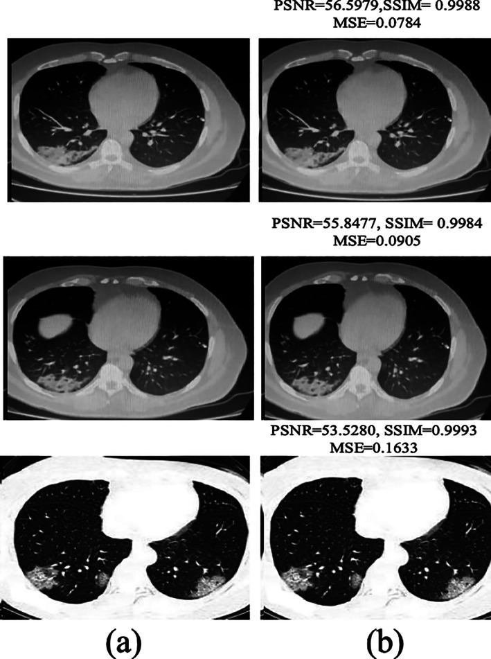
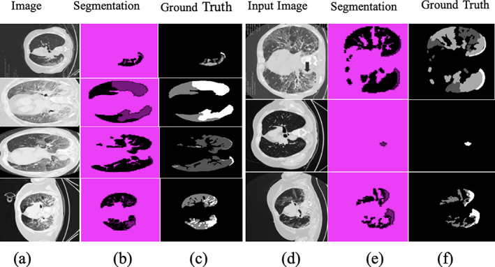
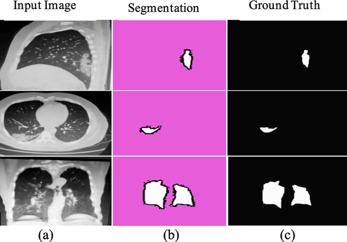
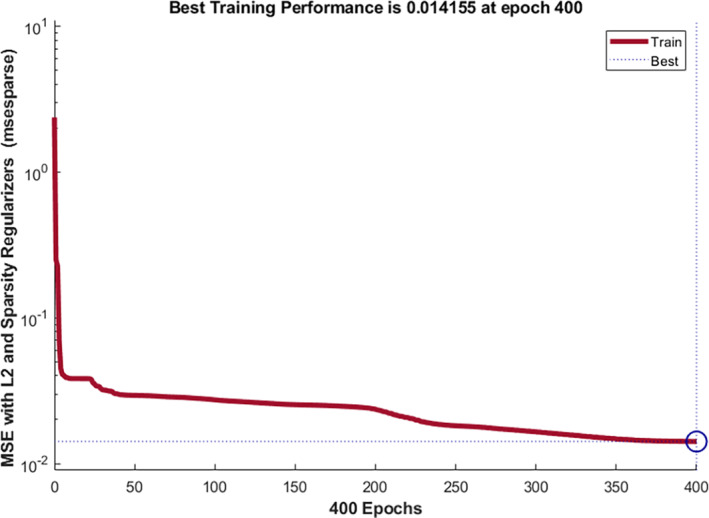
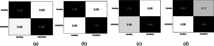
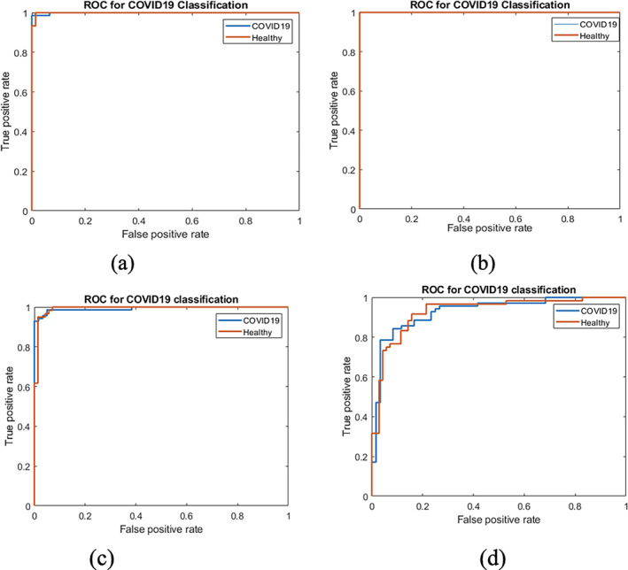
The detection of biological RNA from sputum has a comparatively poor positive rate in the initial/early stages of discovering COVID-19, as per the World Health Organization. It has a different morphological structure as compared to healthy images, manifested by computer tomography (CT). COVID-19 diagnosis at an early stage can aid in the timely cure of patients, lowering the mortality rate. In this reported research, three-phase model is proposed for COVID-19 detection. In Phase I, noise is removed from CT images using a denoise convolutional neural network (DnCNN). In the Phase II, the actual lesion region is segmented from the enhanced CT images by using deeplabv3 and ResNet-18. In Phase III, segmented images are passed to the stack sparse autoencoder (SSAE) deep learning model having two stack auto-encoders (SAE) with the selected hidden layers. The designed SSAE model is based on both SAE and softmax layers for COVID19 classification. The proposed method is evaluated on actual patient data of Pakistan Ordinance Factories and other public benchmark data sets with different scanners/mediums. The proposed method achieved global segmentation accuracy of 0.96 and 0.97 for classification.
Keywords: Deeplabv3; ResNet-18; denoise convolutional neural network (DnCNN); healthcare; public health; stack sparse autoencoder deep learning model (SSAE).
© 2021 Wiley Periodicals LLC.
Conflict of interest statement
The authors declare that they have no conflicts of interest to report regarding the present study.
Figures
Similar articles
-
An intelligence design for detection and classification of COVID19 using fusion of classical and convolutional neural network and improved microscopic features selection approach.Microsc Res Tech. 2021 Oct;84(10):2254-2267. doi: 10.1002/jemt.23779. Epub 2021 May 8. Microsc Res Tech. 2021. PMID: 33964096 Free PMC article.
-
CAD systems for COVID-19 diagnosis and disease stage classification by segmentation of infected regions from CT images.BMC Bioinformatics. 2022 Jul 6;23(1):264. doi: 10.1186/s12859-022-04818-4. BMC Bioinformatics. 2022. PMID: 35794537 Free PMC article.
-
Deep Learning Algorithm for COVID-19 Classification Using Chest X-Ray Images.Comput Math Methods Med. 2021 Nov 9;2021:9269173. doi: 10.1155/2021/9269173. eCollection 2021. Comput Math Methods Med. 2021. PMID: 34795794 Free PMC article.
-
Classification of COVID-19 patients from chest CT images using multi-objective differential evolution-based convolutional neural networks.Eur J Clin Microbiol Infect Dis. 2020 Jul;39(7):1379-1389. doi: 10.1007/s10096-020-03901-z. Epub 2020 Apr 27. Eur J Clin Microbiol Infect Dis. 2020. PMID: 32337662 Free PMC article.
-
A computer-aided diagnosis system for the classification of COVID-19 and non-COVID-19 pneumonia on chest X-ray images by integrating CNN with sparse autoencoder and feed forward neural network.Comput Biol Med. 2022 Feb;141:105134. doi: 10.1016/j.compbiomed.2021.105134. Epub 2021 Dec 14. Comput Biol Med. 2022. PMID: 34971978 Free PMC article.
Cited by
-
COVID-DAI: A novel framework for COVID-19 detection and infection growth estimation using computed tomography images.Microsc Res Tech. 2022 Jun;85(6):2313-2330. doi: 10.1002/jemt.24088. Epub 2022 Feb 23. Microsc Res Tech. 2022. PMID: 35194866 Free PMC article.
-
D2BOF-COVIDNet: A Framework of Deep Bayesian Optimization and Fusion-Assisted Optimal Deep Features for COVID-19 Classification Using Chest X-ray and MRI Scans.Diagnostics (Basel). 2022 Dec 29;13(1):101. doi: 10.3390/diagnostics13010101. Diagnostics (Basel). 2022. PMID: 36611393 Free PMC article.
-
Three-Dimensional Semantic Segmentation of Diabetic Retinopathy Lesions and Grading Using Transfer Learning.J Pers Med. 2022 Sep 5;12(9):1454. doi: 10.3390/jpm12091454. J Pers Med. 2022. PMID: 36143239 Free PMC article.
-
Computed Tomography Image Features under Denoising Algorithm for Benign and Malignant Diagnosis of Renal Parenchymal Tumor.Contrast Media Mol Imaging. 2022 May 27;2022:5871385. doi: 10.1155/2022/5871385. eCollection 2022. Contrast Media Mol Imaging. 2022. PMID: 35685673 Free PMC article.
-
Liver Tumor Localization Based on YOLOv3 and 3D-Semantic Segmentation Using Deep Neural Networks.Diagnostics (Basel). 2022 Mar 27;12(4):823. doi: 10.3390/diagnostics12040823. Diagnostics (Basel). 2022. PMID: 35453870 Free PMC article.
References
-
- Amin, J. , Sharif, M. , Raza, M. , Saba, T. , & Anjum, M. A. (2019). Brain tumor detection using statistical and machine learning method. Computer Methods and Programs in Biomedicine, 177, 69–79. - PubMed
-
- Amin, J. , Sharif, M. , Raza, M. , Saba, T. , & Rehman, A. (2019). Brain tumor classification: feature fusion. 2019 International Conference on Computer and Information Sciences (ICCIS) (pp. 1‐6). IEEE.
-
- Amin, J. , Sharif, M. , Raza, M. , Saba, T. , Sial, R. , & Shad, S. A. (2020). Brain tumor detection: A long short‐term memory (LSTM)‐based learning model. Neural Computing and Applications, 32, 15965–15973.
-
- Amin, J. , Sharif, M. , Yasmin, M. , Saba, T. , & Raza, M. (2019). Use of machine intelligence to conduct analysis of human brain data for detection of abnormalities in its cognitive functions. Multimedia Tools & Applications, 79(15), 10955–10973. 10.1007/s11042-019-7324-y - DOI
MeSH terms
LinkOut - more resources
Full Text Sources
Medical
