. 2021 Aug 12;7(8):653.
doi: 10.3390/jof7080653.

Basidiobolus omanensis sp. nov. Causing Angioinvasive Abdominal Basidiobolomycosis

Affiliations

Basidiobolus omanensis sp. nov. Causing Angioinvasive Abdominal Basidiobolomycosis

Abdullah M S Al-Hatmi et al. J Fungi (Basel). .

Abstract

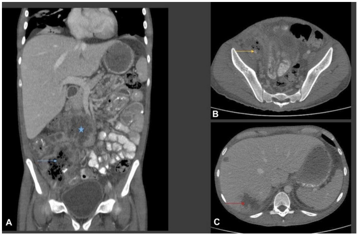
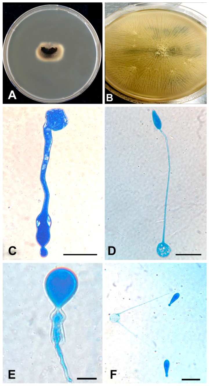
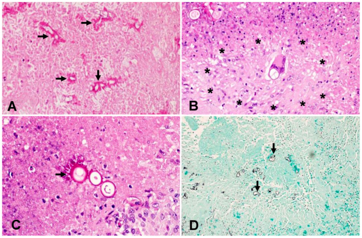
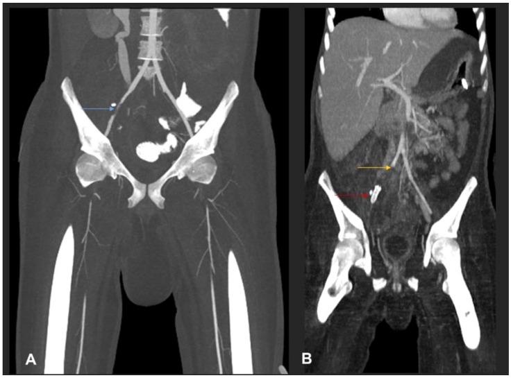
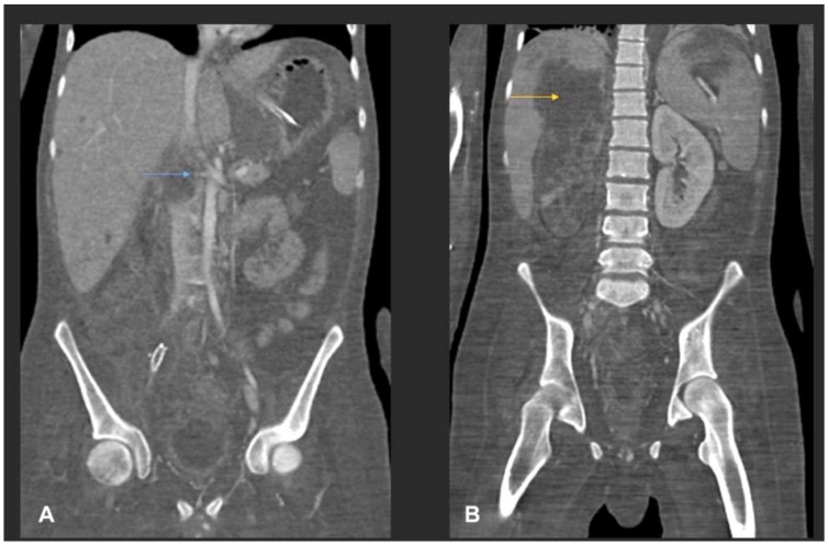
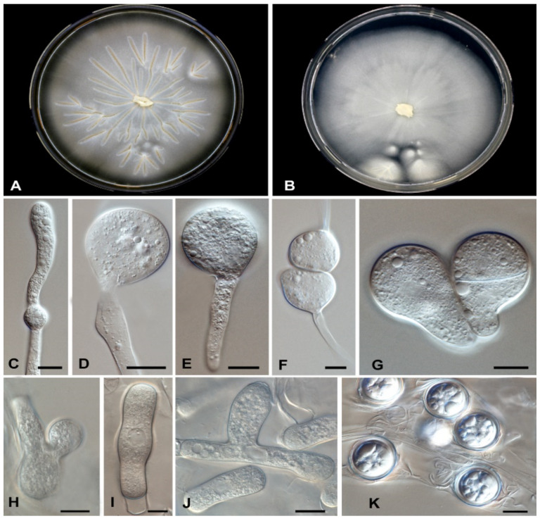
Human infectious fungal diseases are increasing, despite improved hygienic conditions. We present a case of gastrointestinal basidiobolomycosis (GIB) in a 20-year-old male with a history of progressively worsening abdominal pain. The causative agent was identified as a novel Basidiobolus species. Validation of its novelty was established by analysis of the partial ribosomal operon of two isolates from different organs. Phylogeny of ITS and LSU rRNA showed that these isolates belonged to the genus Basidiobolus, positioned closely to B. heterosporus and B. minor. Morphological and physiological data supported the identity of the species, which was named Basidiobolus omanensis, with CBS 146281 as the holotype. The strains showed high minimum inhibitory concentrations (MICs) to fluconazole (>64 µg/mL), itraconazole and voriconazole (>16 µg/mL), anidulafungin and micafungin (>16 µg/mL), but had a low MIC to amphotericin B (1 µg/mL). The pathogenic role of B. omanensis in gastrointestinal disease is discussed. We highlight the crucial role of molecular identification of these rarely encountered opportunistic fungi.

Keywords: Basidiobolus; ITS; LSU; basidiobolomycosis; gastrointestinal; morphology; phylogeny.


Conflict of interest statement

The authors declare no conflict of interest.

Figures

Figure 1
Figure 1
Multiplanar CT scan of abdomen with oral and intravenous contrast. (A) Coronal CT image shows a multispatial ill-defined low-attenuation ring enhancing collection in the root of the mesentery (star) and in the right iliac fossa. The right iliac fossa collection shows thick irregular rim enhancement with central bubbly air lucency (blue arrow). Bowel loops are adherent to the collections. There is marked inflammatory fat stranding noted in the right iliac fossa and right side of the pelvic cavity. Small volume ascites. (B) Axial CT image from the right iliac fossa and pelvis shows marked phlegmonous soft tissue lesion with fat stranding. The phlegmonous inflammatory mass is encasing the right external iliac artery which is small and irregular in calibre (yellow arrow). (C) Axial images from the liver show multifocal ill-defined low-attenuation liver lesions of variable size (two of them shown in this image). The largest was seen in the subcapsular liver in the segment VII and shows surround oedema, suggestive of liver abscess (red arrow).
Figure 2
Figure 2
(A) Direct culture (SAB) from the thrombus. A piece of the thrombus (the black material at the centre) was put on a SAB plate. After incubation, the mould grew from it. (B) Colonial appearance of flat, radially folded, waxy, yellow-cream colonies on SAB. (CE) Wide hyphae and club-shaped spores with knob-like tips demonstrated with lactophenol cotton blue stain. (F) The thin ballistoconidia in the primary culture. All scale 10 µm.
Figure 3
Figure 3
Histopathology findings of a section of small intestine: (A) HE stain (4×), within the necrotic areas, there are numerous fungal elements (arrows) invading all the layers beyond the mucosa. The fungi are characterised by thin walls and broad septate hyphae in keeping with basidiobolomycosis. (B) HE stain (40×), the fungal hyphae are associated with dense fibro-inflammatory reaction with numerous necrotising granulomas. In this image, a granuloma’s border is marked by asterisks. (C) HE stain (40×), hyphae are frequently surrounded by eosinophilic material known as Splendore–Hoeppli phenomenon (arrow). (D) GMS stain (20×), the fungi appear black with GMS stain (arrows). HE: haematoxylin and eosin; GMS: Gomori methenamine silver.
Figure 4
Figure 4
(A) MIP coronal CT angiography of aorta and lower limb shows a segmental narrowing and luminal irregularities of right external iliac artery (blue arrow). No occlusion. Distal run off was satisfactory. Rest of the lower limb vessels were patent and normal (not shown). (B) MIP coronal image of the aorta and lower limbs shows cut-off of the proximal right common iliac artery suggestive of thrombotic occlusion (yellow arrow). A stent is seen in the right external iliac artery (red arrow). Right iliac fossa shows extensive inflammatory phlegmonous mass-like soft tissue thickening.
Figure 5
Figure 5
(A) MIP coronal images of abdomen shows a cut-off of the right proximal renal artery 1 cm from the ostium with adjacent inflammatory collection (blue arrow). (B) Coronal post-contrast CT shows infarction of the right kidney. A large liver abscess (yellow arrow) in the right liver lobe with extracapsular extension into the upper pole of the right kidney is also noted.
Figure 6
Figure 6
Maximum Likelihood (RAxML) phylogram obtained from the combined analysis of ITS and LSU sequences of Basidiobolus spp. Numbers on the nodes are Bayesian posterior probability values (BI-PP) ≥ 0.95, followed by ML bootstrap values (BS) ≥ 70%. Novel taxa proposed in this study are indicated in bold. Ex-type strains are indicated with T. The tree was rooted to Conidiobolus sp. (ARSEF 7942).
Figure 7
Figure 7
Basidiobolus omanensis. A colony on (A) MEA, (B) SAB after 3–5 days at 25 °C, (C) mycelia with young chlamydospore, (D) primary conidiophore arising from mycelia, (E) primary conidia, (F) formation of zygospores, (G) mature septate conidia, (H) mature zygospores, (I,J) branched and unbranched hyphae, (K) mature zygospores. Bars 10 μm.

References

    1. Joe L.K., Njo-Injo T.E., Pohan A., Van Der Meulen H. Basidiobolus ranarum as a cause of subcutaneous phycomycosis in Indonesia. Arch. Dermatol. Syphol. 1956;74:378–383. doi: 10.1001/archderm.1956.01550100046008. - DOI - PubMed
    1. Van Overeem C. Uber ein merkwurdiges Vorkommen von Basidiobolus ranarum Eidam. Bull. Jardin Bot. 1925;7:423–431.
    1. Greene C.E., Brockus C.W., Currin M.P., Jones C.J. Infection with Basidiobolus ranarum in two dogs. J. Am. Vet. Med. Assoc. 2002;221:528–532. doi: 10.2460/javma.2002.221.528. - DOI - PubMed
    1. Al-Maani A.S., Paul G., Jardani A., Nayar M., Al-Lawati F., Al-Baluishi S., Hussain I.B. Gastrointestinal basidiobolomycosis: First case report from Oman and literature review. Sultan Qaboos Univ. Med. J. 2014;14:e241. - PMC - PubMed
    1. Khan Z.U., Khoursheed M., Makar R., Al-Waheeb S., Al-Bader I., Al-Muzaini A., Chandy R., Mustafa A.S. Basidiobolus ranarum as an etiologic agent of gastrointestinal zygomycosis. J. Clin. Microbiol. 2001;39:2360–2363. doi: 10.1128/JCM.39.6.2360-2363.2001. - DOI - PMC - PubMed

LinkOut - more resources