Review
. 2021 Jul 30;8(8):89.
doi: 10.3390/jcdd8080089.

The Cardiac Neural Crest Cells in Heart Development and Congenital Heart Defects

Affiliations
Review

The Cardiac Neural Crest Cells in Heart Development and Congenital Heart Defects

Shannon Erhardt et al. J Cardiovasc Dev Dis. .

Abstract

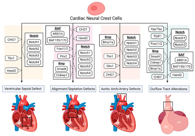
The neural crest (NC) is a multipotent and temporarily migratory cell population stemming from the dorsal neural tube during vertebrate embryogenesis. Cardiac neural crest cells (NCCs), a specified subpopulation of the NC, are vital for normal cardiovascular development, as they significantly contribute to the pharyngeal arch arteries, the developing cardiac outflow tract (OFT), cardiac valves, and interventricular septum. Various signaling pathways are shown to orchestrate the proper migration, compaction, and differentiation of cardiac NCCs during cardiovascular development. Any loss or dysregulation of signaling pathways in cardiac NCCs can lead to abnormal cardiovascular development during embryogenesis, resulting in abnormalities categorized as congenital heart defects (CHDs). This review focuses on the contributions of cardiac NCCs to cardiovascular formation, discusses cardiac defects caused by a disruption of various regulatory factors, and summarizes the role of multiple signaling pathways during embryonic development. A better understanding of the cardiac NC and its vast regulatory network will provide a deeper insight into the mechanisms of the associated abnormalities, leading to potential therapeutic advancements.

Keywords: cardiac neural crest; congenital heart defects (CHDs); heart development; neural crest cells (NCCs); outflow tract (OFT).


Conflict of interest statement

The authors declare no conflict of interest.

Figures

Figure 1
Figure 1
The disruption of various genes and signaling pathways, indicated to be important for proper cardiac neural crest (NC) contribution, results in numerous congenital heart defect (CHD) phenotypes. Such phenotypes include ventricular septal defect (VSD), various vascular defects including misalignment of the aorta and pulmonary trunks and aortic artery abnormalities, and outflow tract (OFT) alterations, such as OFT constriction and improper septation. Loss of Tbx1 in mice has been shown to alter the environment of migrating neural crest cells (NCCs), producing great vessel abnormalities and aortic arch interruption, while also presenting with VSD in patients. CHD7 mutations within NCCs can produce septum defects such as VSD, and vascular alterations such as double outlet right ventricle (DORV) and interrupted aortic arch, along with inhibiting NC contributions to the OFT. In NCCs, disrupted Notch2 results in a narrowed OFT and OFT arteries, while mutations of all Notch-target genes (Notch1, Notch2, Notch3, Notch4) have been shown to result in VSD, along with vascular abnormalities such as pulmonic stenosis and aortic arch patterning defects. The deletion of a known bone morphogenetic protein (Bmp) inhibitor, Ctdnep1, produced premature and asymmetric OFT septation, pulmonary closure, premature NCC compaction, and embryonic death, while the loss of the Bmp receptor Bmpr1a, resulted in shortened OFTs leading to persistent truncus arteriosus (PTA), along with occasional aortic arch abnormalities. It was also found that the deletion of Smad4, located downstream of the Bmp network, also resulted in shortened OFTs, along with some hearts presenting with no OFT septation, resulting in PTA. The ablation of Yap and Taz, key components of the Hippo signaling pathway, in NCCs have been shown to disrupt smooth muscle expression and differentiation of the OFT. Deletions of BAF155 and BAF170 in NCCs have resulted in impaired NCC contribution for OFT development, while a loss of ARID1A, a BAF (Brg1/Brm-associated factors) subunit, has shown improper NC contribution for OFT development with producing conotruncal septation defects. The disruption of Pitx2 and Pitx2 upstream regulators suggests a negative impact on cardiac NC abilities for proper vasculature development. Hand2 deletions in NCCs present with a large variety of altered cardiac morphology, such as impaired OFT development, DORV, and VSD. Lack of OFT septation is also seen with the deletion of TGIF1 in NCCs. Deletion of Foxc1/2 in mice presents with lacking or shortened OFTs, along with improper OFT septation and PTA. Ao, aorta; Pv, pulmonary vessel; LV, left ventricle; RV, right ventricle; OFT, outflow tract. Created with Biorender.com (accessed on 28 July 2021).

References

    1. Xi M., Lui F. StatPearls. StatPearls Publishing; Treasure Island, FL, USA: 2021. Neuroanatomy, Neural Crest. - PubMed
    1. Sauka-Spengler T., Bronner-Fraser M. A gene regulatory network orchestrates neural crest formation. Nat. Rev. Mol. Cell Biol. 2008;9:557–568. doi: 10.1038/nrm2428. - DOI - PubMed
    1. Arima Y., Miyagawa-Tomita S., Maeda K., Asai R., Seya D., Minoux M., Rijli F.M., Nishiyama K., Kim K.S., Uchijima Y., et al. Preotic neural crest cells contribute to coronary artery smooth muscle involving endothelin signalling. Nat. Commun. 2012;3:1267. doi: 10.1038/ncomms2258. - DOI - PubMed
    1. Etchevers H.C., Dupin E., Le Douarin N.M. The diverse neural crest: From embryology to human pathology. Development. 2019;146 doi: 10.1242/dev.169821. - DOI - PubMed
    1. Rocha M., Beiriger A., Kushkowski E.E., Miyashita T., Singh N., Venkataraman V., Prince V.E. From head to tail: Regionalization of the neural crest. Development. 2020;147 doi: 10.1242/dev.193888. - DOI - PMC - PubMed

LinkOut - more resources