. 2021 Aug 4;8(8):93.
doi: 10.3390/jcdd8080093.

Applicability of the Leiden Convention and the Lipton Classification in Patients with a Single Coronary Artery in the Setting of Congenital Heart Disease

Affiliations

Applicability of the Leiden Convention and the Lipton Classification in Patients with a Single Coronary Artery in the Setting of Congenital Heart Disease

Diana Isabel Katekaru-Tokeshi et al. J Cardiovasc Dev Dis. .

Abstract

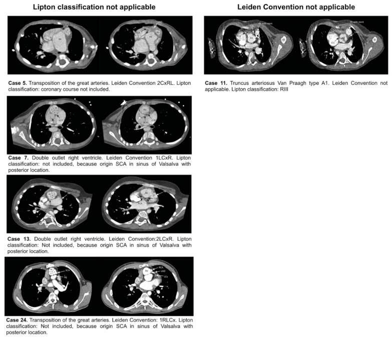
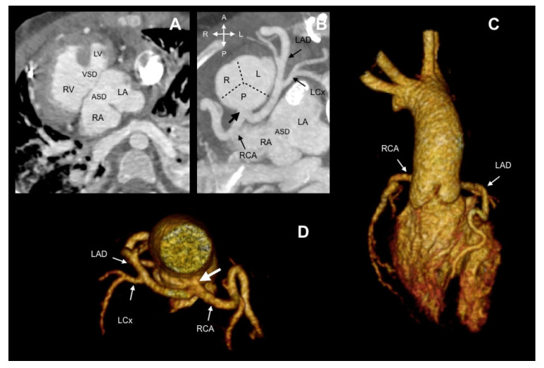
In single coronary artery (SCA) anatomy, all coronary tributaries arise from a single ostium, providing perfusion to the entire myocardium. Coronary classification systems can facilitate the description of SCA anatomy. Aim: Evaluation of the applicability of Lipton classification and the Leiden Convention coronary coding system in SCA. Methods: All patients (n = 6209) who underwent computed tomography (CT) scanning between 2014 and 2018 were retrospectively examined for the presence of SCA and classified, according to Lipton classification and the Leiden Convention coronary coding system. Results: The prevalence of SCA was 0.51% (32/6209). Twenty-eight patients (87.5%) had coexisting congenital heart disease (CHD), most frequently pulmonary atresia (9/32, 28.1%). Ten patients (10/32, 31.25%) could not be classified with either the Leiden Convention or Lipton classification (pulmonary atresia n = 9, common arterial trunk (CAT) n = 1). In one case with CAT, Lipton classification, but not the Leiden Convention, could be applied. In two cases with the transposition of the great arteries and in two cases of double outlet right ventricle, the Leiden Convention, but not the Lipton classification, could be applied. Conclusions: Both classifications are useful to detail information about SCA. As Lipton classification was not developed for structural heart disease cases, in complex CHD with abnormal position of the great arteries, the Leiden Convention is better applicable. The use of both systems is limited in pulmonary atresia. In this scenario, it is better to provide a precise description of the coronary origin and associated characteristics that might affect treatment and prognosis.

Keywords: Leiden Convention coronary coding system; Lipton classification; congenital heart disease; coronary artery anatomy; single coronary artery.


Conflict of interest statement

All authors declare no conflict of interest. This research did not receive any specific grant from funding agencies in the public, commercial, or not-for-profit sectors.

Figures

Figure 1
Figure 1
Classification of SCA by Lipton and modified by Yamanaka and Hobbs (marked). Viewed from caudal to cranial, “imaging view”. Ao: Aorta. L: Left sinus of Valsalva. LAD: Left anterior descending artery. LCx: Left circumflex artery. PA: Pulmonary artery. R: Right sinus of Valsalva. RCA: Right coronary artery.
Figure 2
Figure 2
The Leiden Convention considers a surgical/interventional or an imaging perspective (Figure derived with permission from Koppel et al. [6]). (A) Surgical/Interventional Leiden Convention. In this method, the coronary anatomy is examined from above, as a surgeon would see it during surgery. The physician sits in the non-facing sinus, facing the pulmonary valve. In that position, the sinus on the right is sinus 1 and the sinus on the left is sinus 2. Starting from sinus 1, the coronary branches are named in the order that they are encountered when following a counterclockwise rotation. (B) Imaging Leiden Convention. The physician’s view on the coronary anatomy is from the base of the aorta upward. The physician sits in the non-facing sinus of the aortic valve, facing outward from the sinus. From this position, the right-hand sinus is again sinus 1, and the left-hand sinus is sinus 2. Following a clockwise rotation, starting at sinus 1, the encountered coronary branches are annotated. A: Anterior. Ao: Aorta. L: Left. NF: Non-facing sinus. P: Posterior. Pu: Pulmonary. R: Right.
Figure 3
Figure 3
Case 27. CCTA in a 37-year-old woman undergoing preoperative evaluation for an atrial septal defect. (A) Axial image depicting the origin of the SCA (arrow) from sinus 2. (B) A 3D volume-rendered image showing that the RCA originates from the middle segment of the LAD with an interarterial course. According to the Imaging Leiden Convention, the anatomy is 2R*LCx; by Lipton classification, it is LIIB. (C,D) The RCA follows a course between the aorta and pulmonary artery (level of right ventricular outflow tract). Ao: Aorta. LAD: Left anterior descending coronary artery. LCx: Left circumflex. NF: Non-facing sinus. PA: Pulmonary artery. RCA: Right coronary artery.
Figure 4
Figure 4
Case 9. A 4-year-old child with tetralogy of Fallot. (A) Oblique image showing infundibular pulmonary stenosis (*). (B) Axial image reveals coronary origins clockwise rotated along with the aortic root. According to the Leiden Convention, it is 2RLCx and by Lipton classification, it is LIIA. (C) A 3D volume-rendered image shows an anomalous origin of the RCA from the LAD. A: Anterior. Ao: Aorta. LAD: Left anterior descending coronary artery. L: Left. LCx: Left circumflex. P: Posterior. R: Right. RV: Right ventricle. NF: Non-facing sinus. PA: Pulmonary artery. RCA: Right coronary artery.
Figure 5
Figure 5
Overview of cases that could be classified only with the Lipton classification or the Leiden Convention. Ao: Aorta. LAD: Left anterior descending coronary artery. LCx: Left circumflex. R: Right. NF: Non-facing sinus. PA: Pulmonary artery. RCA: Right coronary artery. SCA: Single coronary artery.
Figure 6
Figure 6
Case 17. A 10-year-old child with pulmonary atresia and common atrioventricular canal type C of Rastelli. (A) Oblique axial view showing atrial situs inversus, common atrioventricular connection, atrial and ventricular septal defect. (B) The SCA (thick arrow) arises from the posterior sinus, which gives origin to the RCA and left main stem. (C,D) A 3D volume-rendered image showing a posterior SCA. Ao: Aorta. ASD: Atrial septal defect. LA: Left atrium. LAD: Left anterior descending coronary artery. LCx. Left circumflex. LV: Left ventricle. NF: Non-facing sinus. RA: Right atrium. RCA: Right coronary artery. RV: Right ventricle. VSD: Ventricular septal defect.

References

    1. Aldana-Sepulveda N., Restrepo C.S., Kimura-Hayama E. Single coronary artery: Spectrum of imaging findings with multidetector CT. J. Cardiovasc. Comput. Tomogr. 2013;7:391–399. doi: 10.1016/j.jcct.2013.11.009. - DOI - PubMed
    1. Lipton M.J., Barry W.H., Obrez I., Silverman J.F., Wexler L. Isolated single coronary artery: Diagnosis, angiographic classification, and clinical significance. Radiology. 1979;130:39–47. doi: 10.1148/130.1.39. - DOI - PubMed
    1. Yamanaka O., Hobbs R.E. Coronary artery anomalies in 126,595 patients undergoing coronary arteriography. Catheter. Cardiovasc. Diagn. 1990;21:28–40. doi: 10.1002/ccd.1810210110. - DOI - PubMed
    1. Gittenberger-de Groot A.C., Sauer U., Oppenheimer-Dekker A., Quaegebeur J. Coronary arterial anatomy in transposition of the great arteries: A morphologic study. Pediatr. Cardiol. 1983;4:15–24.
    1. Gittenberger-de Groot A.C., Koenraadt W.M.C., Bartelings M.M., Bökenkamp R., DeRuiter M.C., Hazekamp M.G., Bogers A., Quaegebeur J.M., Schalij M.J., Vliegen H.W., et al. Coding of coronary arterial origin and branching in congenital heart disease: The modified Leiden Convention. J. Thorac. Cardiovasc. Surg. 2018;156:2260–2269. doi: 10.1016/j.jtcvs.2018.08.009. - DOI - PubMed