. 2021 Oct 1;139(10):1096-1103.
doi: 10.1001/jamaophthalmol.2021.3303.

Continued Increase of Axial Length and Its Risk Factors in Adults With High Myopia

Affiliations

Continued Increase of Axial Length and Its Risk Factors in Adults With High Myopia

Ran Du et al. JAMA Ophthalmol. .

Abstract

Importance: Pathologic myopia due to an excessive increase of axial length is associated with severe visual impairments. Systematic analyses to determine the rate of and the risk factors associated with the axial elongation in adults with high myopia based on long-term follow-up of a large population are needed.

Objective: To determine the risk factors associated with axial elongation in adults with high myopia.

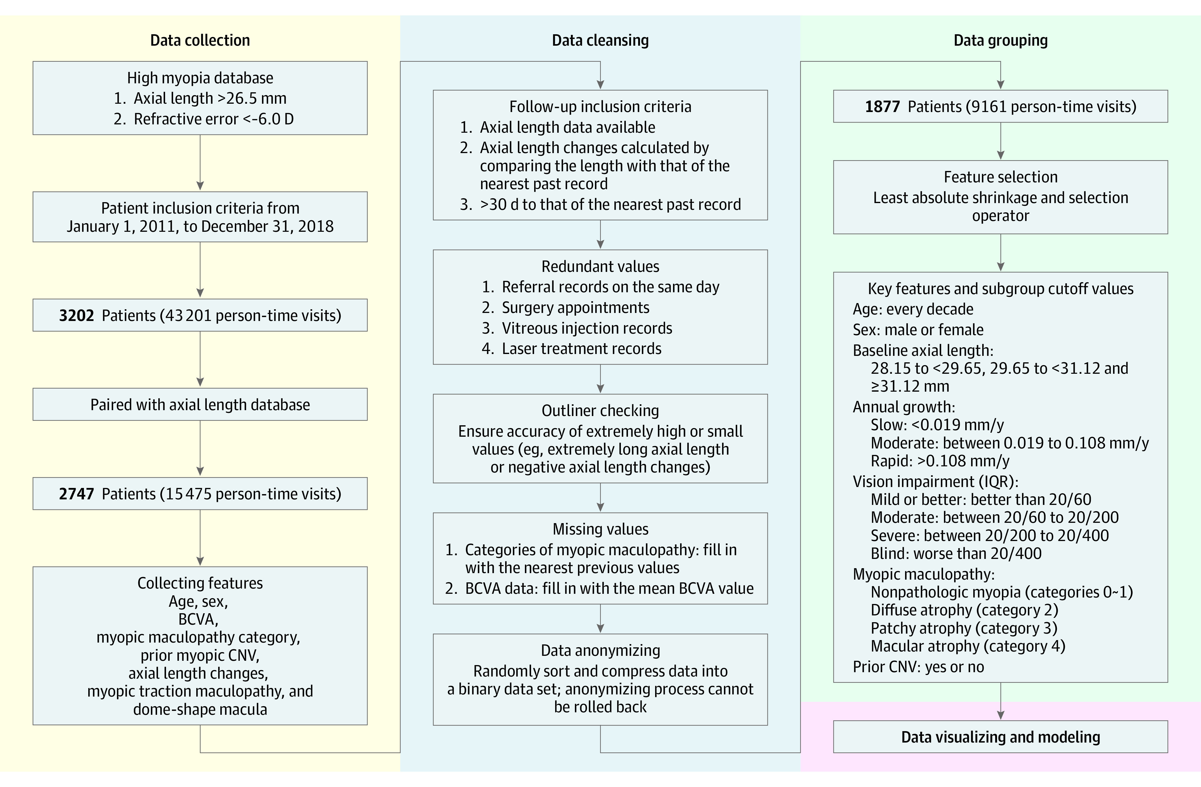
Design, setting, and participants: This cohort study used the medical records of 43 201 patient visits in a single-hospital database that were collected from January 3, 2011, to December 28, 2018. A total of 15 745 medical records with the patients' sex, best-corrected visual acuity (BCVA), axial length, type of myopic maculopathy, and the presence or absence of choroidal neovascularization (CNV) were reviewed. Data were analyzed from April 3, 2019, to August 5, 2020.

Main outcomes and measures: Changes in the axial length at each examination were calculated. The significance of the associations between the annual increase of the axial length and age, sex, baseline axial length, types of myopic maculopathy, and a history of CNV was determined. Generalized linear mixed models were used to evaluate the strength of the risk factors associated with an increase of the axial length in high myopia.

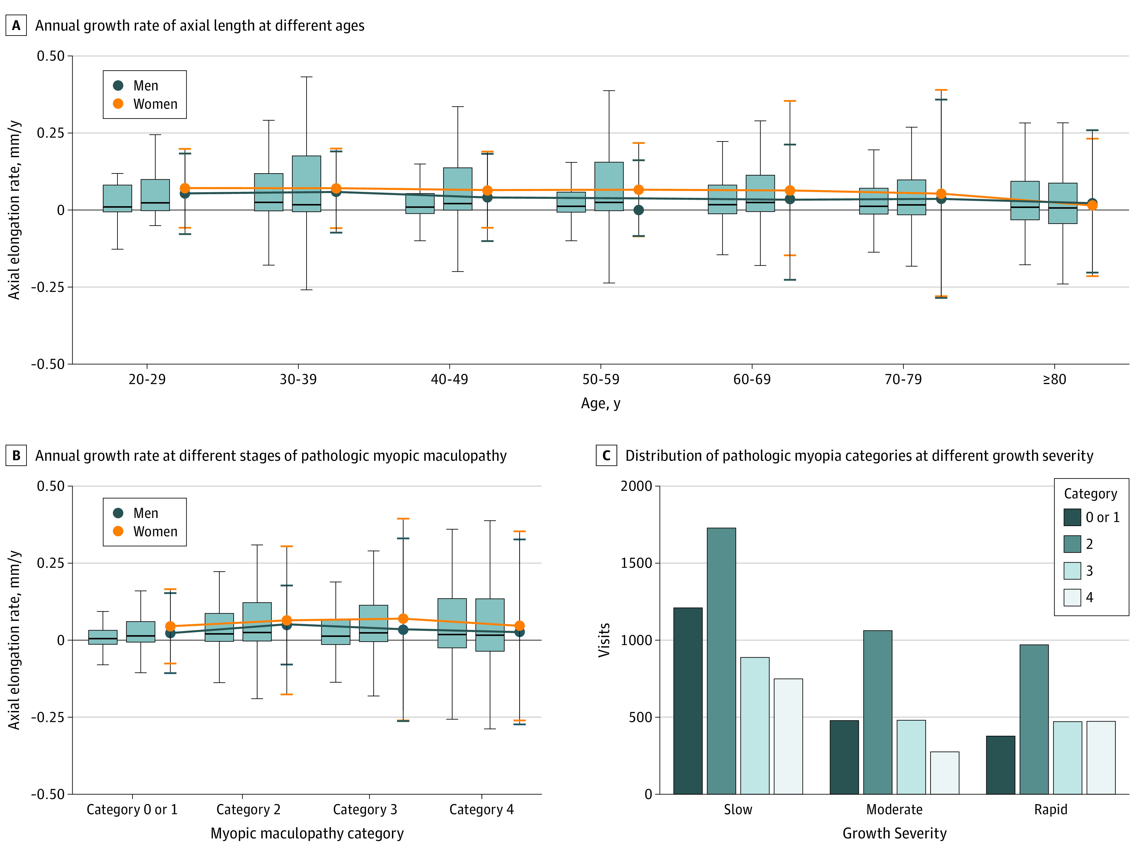
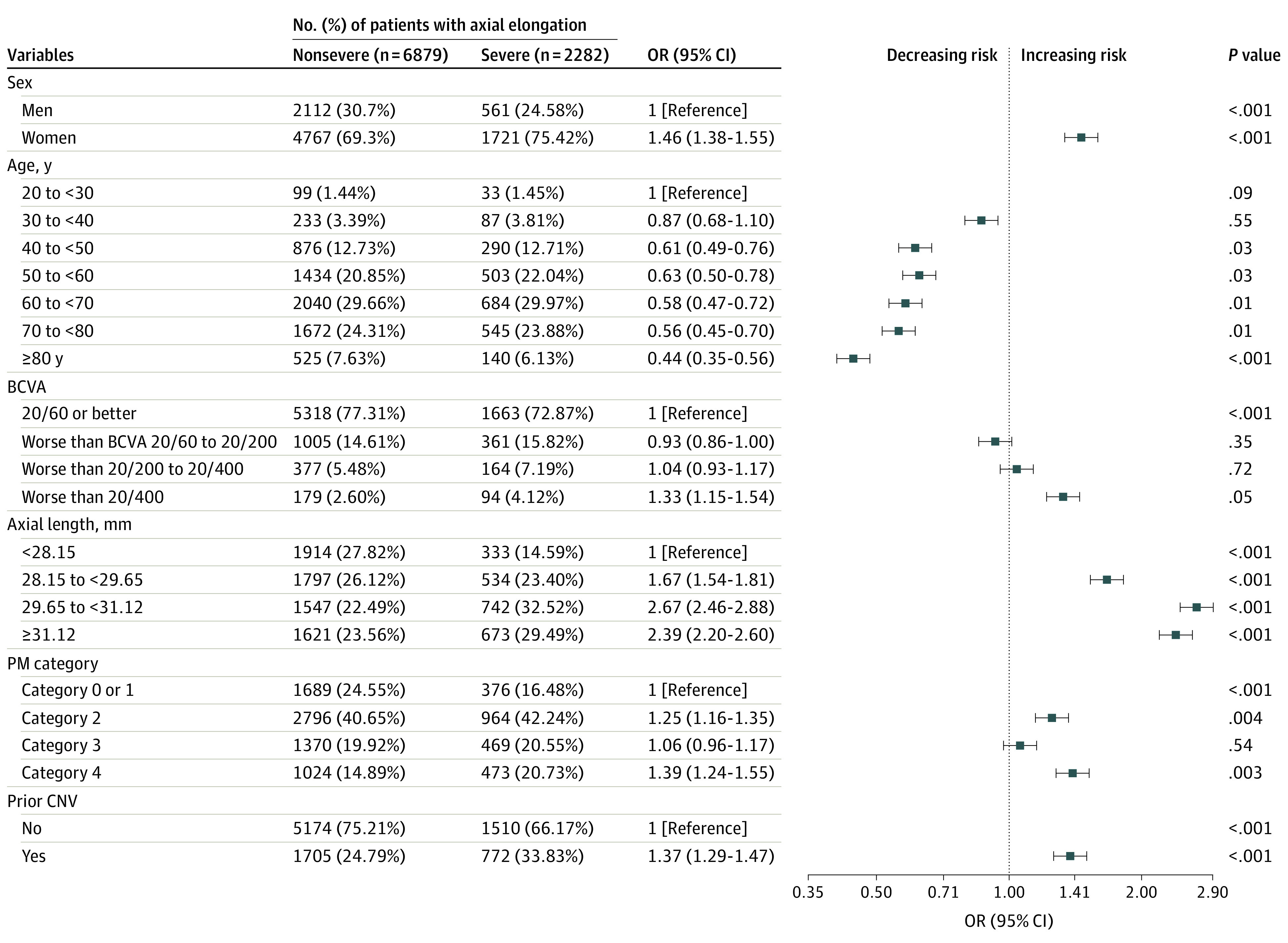
Results: Among 1877 patients with 9161 visits included in the analysis, the mean (SD) age was 62.10 (12.92) years, and 1357 (72.30%) were women. The mean (SD) axial length was 29.66 (2.20) mm with a mean (SD) growth rate of 0.05 (0.24) mm/y. Among the 9161 visits, 7096 eyes (77.46%) had myopic maculopathy and 2477 eyes (27.04%) had CNV. The odds ratio for inducing a severe elongation of the axial length was 1.46 (95% CI, 1.38-1.55) for female sex, 0.44 (95% CI, 0.35-0.56) to 0.63 (95% CI, 13 0.50-0.78) for older than 40 years, 1.33 (95% CI, 1.15-1.54) for BCVA of less than 20/400, 1.67 (95% CI, 1.54-1.81) to 2.67 (95% CI, 2.46-2.88) for baseline axial length of 28.15 mm or greater, 1.06 (95% CI, 0.96-1.17) to 1.39 (95% CI, 1.24-1.55) for the presence of maculopathy, and 1.37 (95% CI, 1.29-1.47) for prior CNV.

Conclusions and relevance: This cohort study found continuing axial elongation in adults with high myopia. The risk factors for elongation do not appear to be modifiable, so prevention of myopia may be the best approach to reduce the incidence of pathologic myopia and its complications in the future.


Conflict of interest statement

Conflict of Interest Disclosures: None reported.

Figures

Figure 1.
Figure 1.. Schematic of Workflow
Our project consisted of 4 steps: data collection, data cleansing, data grouping, and data display. First, we collected medical records that included basic information about patient sex, age, and other relevant data. Then, data cleansing was performed, with a particular focus on filtering out redundant values and inserting missing values. The data were then grouped by different criteria, analyzed, and displayed. BCVA indicates best-corrected visual acuity; CNV, choroidal neovascularization; IQR, interquartile range.
Figure 2.
Figure 2.. Plots of Risk Factors Associated With Axial Elongation
A, The rate of axial elongation is slightly slower after 30 years of age. The mean annual growth of axial length among women is higher than that among men. Box plots represent means and SDs (error bars indicate 95% CIs); dot plots represent medians and interquartile ranges. For each subgroup, the left box plot represents men and the right box plot represents women. B, The annual growth rate was greater in eyes with pathologic myopia, including those with diffuse atrophy (category 2), patchy atrophy (category 3), and macular atrophy (category 4) (maculopathy) than eyes with nonpathologic myopia (category 0) or tessellated fundus (category 1) among men. Among women, the annual growth rate was greater in eyes with categories 2 and 3 maculopathy than eyes with nonpathologic myopia, and no significant difference was seen in the growth rate between women with category 4 or with categories 0 or 1. The annual growth rate among women was greater in eyes with categories 2 (P = .008) and 3 (P < .001) maculopathy compared with that among men. Box plots represent means and SDs (error bars indicate 95% CIs); dot plots represent medians and interquartile ranges. For each subgroup, the left box plot represents men and the right box plot represents women. C, Proportions of eyes with categories 3 and 4 myopic maculopathy in the group with a moderate increase in axial length (0.019 to <0.108 mm) and in the rapid increase group (≥0.108 mm) are compared with the slow increase group (<0.019 mm).
Figure 3.
Figure 3.. Odds Ratios (ORs) of Risk Factors Between Groups With and Without Severe Axial Elongation
Severe axial elongation was 0.108 mm/y or greater; nonsevere, less than 0.108 mm/y. Factors were categorized according to each grouping criteria and regressed by using logistic regression to test the differences between the continuous variables that are controlled for multiple confounders. BCVA indicates best-corrected visual acuity; CNV, choroidal neovascularization; PM, pathologic myopia.

References

    1. Lee MW, Lee SE, Lim HB, Kim JY. Longitudinal changes in axial length in high myopia: a 4-year prospective study. Br J Ophthalmol. 2020;104(5):600-603. doi:10.1136/bjophthalmol-2019-314619 - DOI - PubMed
    1. World Medical Association . World Medical Association Declaration of Helsinki: ethical principles for medical research involving human subjects. JAMA. 2013;310(20):2191-2194. doi:10.1001/jama.2013.281053 - DOI - PubMed
    1. Ohno-Matsui K, Kawasaki R, Jonas JB, et al. ; META-analysis for Pathologic Myopia (META-PM) Study Group . International photographic classification and grading system for myopic maculopathy. Am J Ophthalmol. 2015;159(5):877-83.e7. doi:10.1016/j.ajo.2015.01.022 - DOI - PubMed
    1. Ohno-Matsui K, Lai TY, Lai CC, Cheung CM. Updates of pathologic myopia. Prog Retin Eye Res. 2016;52:156-187. doi:10.1016/j.preteyeres.2015.12.001 - DOI - PubMed
    1. Allen M, Poggiali D, Whitaker K, Marshall TR, Kievit RA. Raincloud plots: a multi-platform tool for robust data visualization. Wellcome Open Res. 2019;4(63):63. doi:10.12688/wellcomeopenres.15191.1 - DOI - PMC - PubMed

Publication types