Review
. 2021 Nov;39(11):2295-2309.
doi: 10.1002/jor.25172. Epub 2021 Sep 10.

Methodology, selection, and integration of fracture healing assessments in mice

Affiliations
Review

Methodology, selection, and integration of fracture healing assessments in mice

Adam M Knox et al. J Orthop Res. 2021 Nov.

Abstract

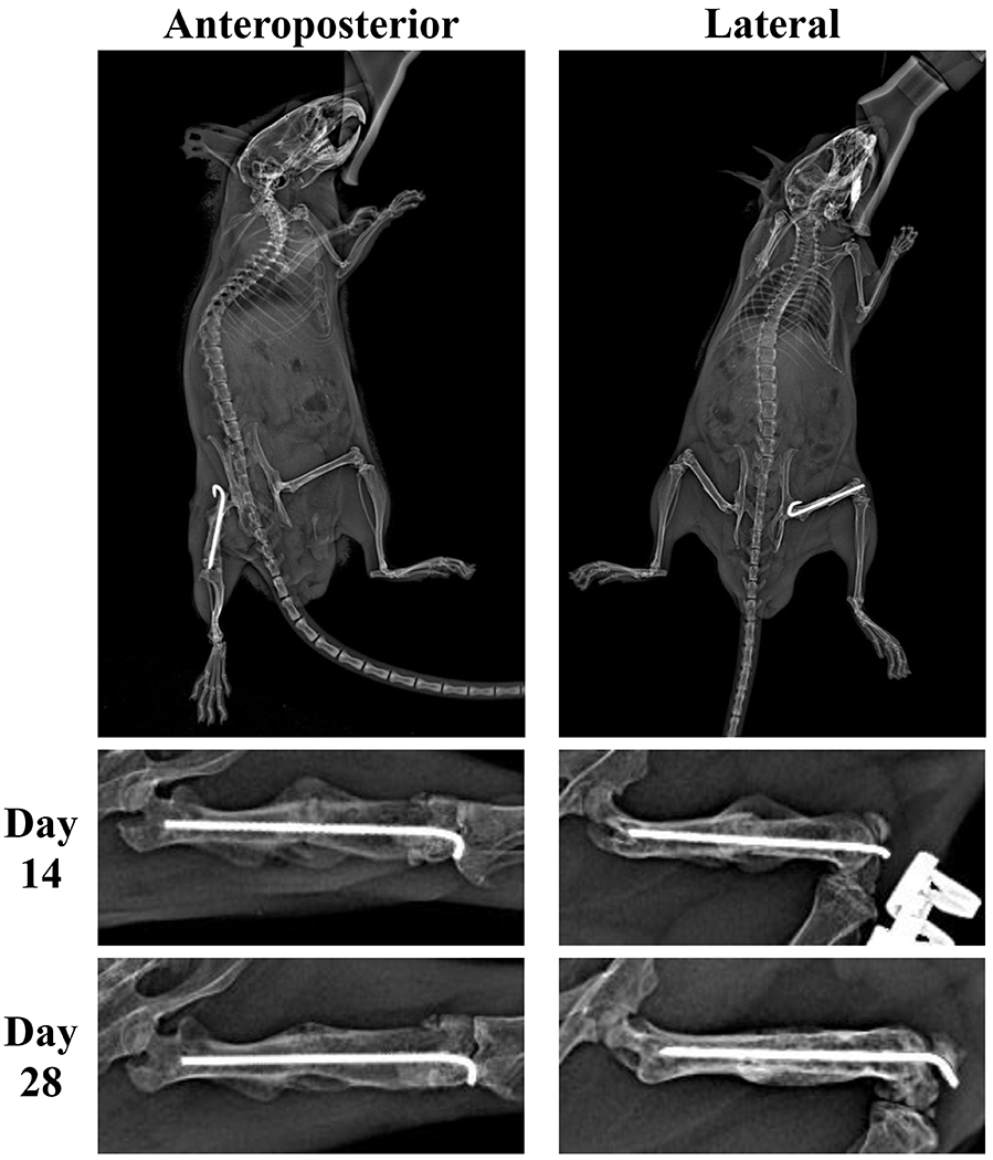
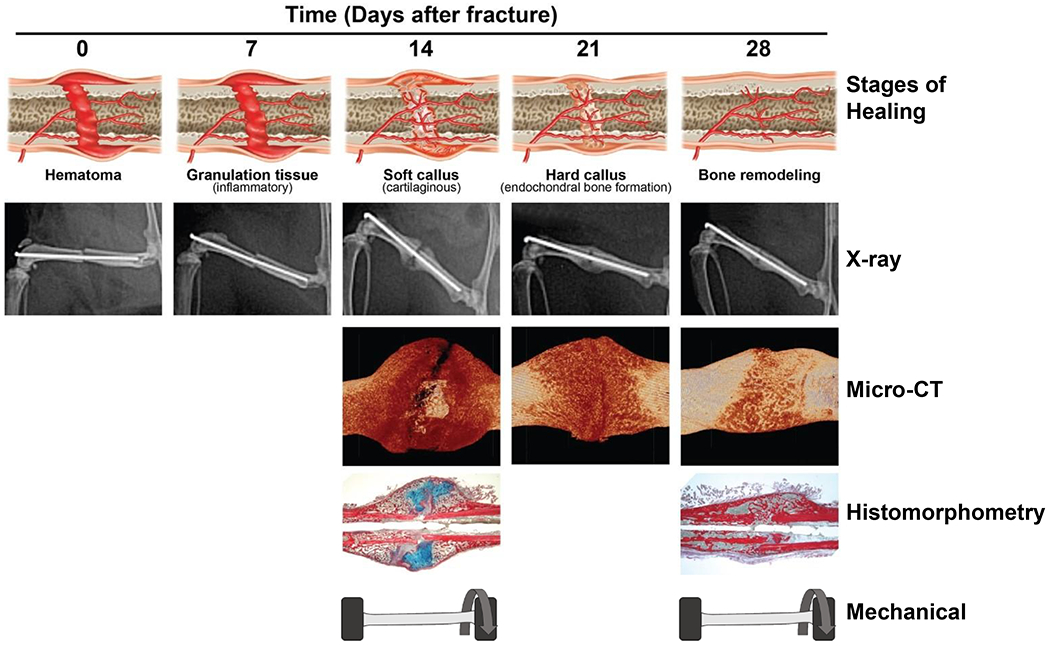
Long bone fractures are one of the most common and costly medical conditions encountered after trauma. Characterization of the biology of fracture healing and development of potential medical interventions generally involves animal models of fracture healing using varying genetic or treatment groups, then analyzing relative repair success via the synthesis of diverse assessment methodologies. Murine models are some of the most widely used given their low cost, wide variety of genetic variants, and rapid breeding and maturation. This review addresses key concerns regarding fracture repair investigations in mice and may serve as a guide in conducting and interpreting such studies. Specifically, this review details the procedures, highlights relevant parameters, and discusses special considerations for the selection and integration of the major modalities used for quantifying fracture repair in such studies, including X-ray, microcomputed tomography, histomorphometric, biomechanical, gene expression and biomarker analyses.

Keywords: X-ray; biomechanics; fracture healing; histology; mRUST; microcomputed tomography; mouse models; preclinical.


Figures

Figure 1.
Figure 1.
Example of radiographic imaging of fracture site from both lateral and anteroposterior views. These two perspectives enable evaluation of callus formation and fracture line at “four cortices” of bone. The cropped images represent longitudinal assessment of fracture healing.
Figure 2.
Figure 2.
Overview of initial steps to generating volume of interest (VOI). Shadow projection image of fractured femur (A). Fracture midline (B ii.) determined by calculating the midpoint between the first intact cortical ring, proximally (B i.) and distally (B iii.) from the fracture. Complete VOI designated 3.5mm above and below fracture line (C-D). Reprinted with permission from Collier et al (15).
Figure 3.
Figure 3.
Overview of binarization and final steps of volume of interest (VOI) selection. Example transverse slices through fracture center after binarization (A) and final VOI selection (B) yielding final mineralized callus model (C-D) for morphometric 3D analysis. Reprinted with permission from Collier et al (15).
Figure 4.
Figure 4.
Representative histologic sections stained with picrosirius red (bone) and alcian blue (cartilage) ready for image processing software analysis. Many alternative histologic staining protocols are available for histomorphometric assessment. Adapted with permission from Collier et al (15).
Figure 5.
Figure 5.
Schematics for biomechanical testing: (A) Three-point bending, (B) Four-point bending, and (C) Torsion testing.
Figure 6.
Figure 6.
Example torque-angular displacement curve generated from torsional testing. Outcome measures include ultimate torque (torque to fracture point), torsional stiffness (relationship between torque and angular displacement prior to the yield point), twist to failure (angular displacement to fracture point) and toughness (total energy to fracture point; area under torque-angular displacement curve).
Figure 7.
Figure 7.
Overview of approximate fracture healing progression and corresponding assessments from 0 to 28 days post fracture.

References

    1. Ensrud KE. Epidemiology of fracture risk with advancing age. J Gerontol A Biol Sci Med Sci. 2013;68(10):1236–42. Epub 2013/07/09. doi: 10.1093/gerona/glt092. - DOI - PubMed
    1. Burge R, Dawson-Hughes B, Solomon DH, et al. Incidence and economic burden of osteoporosis-related fractures in the United States, 2005-2025. J Bone Miner Res. 2007;22(3):465–75. Epub 2006/12/06. doi: 10.1359/jbmr.061113. - DOI - PubMed
    1. Schnell S, Friedman SM, Mendelson DA, et al. The 1-year mortality of patients treated in a hip fracture program for elders. Geriatr Orthop Surg Rehabil. 2010;1(1):6–14. Epub 2010/09/01. doi: 10.1177/2151458510378105. - DOI - PMC - PubMed
    1. Keating JF, O’Brien PJ, Blachut PA, et al. Locking intramedullary nailing with and without reaming for open fractures of the tibial shaft. A prospective, randomized study. J Bone Joint Surg Am. 1997;79(3):334–41. Epub 1997/03/01. doi: 10.2106/00004623-199703000-00003. - DOI - PubMed
    1. group TIw, Busse JW, Bhandari M, et al. Re-evaluation of low intensity pulsed ultrasound in treatment of tibial fractures (TRUST): randomized clinical trial. BMJ. 2016;355:i5351. Epub 2016/11/01. doi: 10.1136/bmj.i5351. at www.icmje.org/coi_disclosure.pdf - DOI - PMC - PubMed
    2. and declare: TAE, ES, and MB have received consulting fees from Smith & Nephew, the manufacturer of the study device. PT receives royalties from Smith & Nephew. GJDR is a paid consultant for Bioventus LLC, which is 51% owned by Essex Woodlands and 49% by Smith & Nephew. MB is supported, in part, by a Canada research chair, McMaster University.

Publication types