. 2021 Aug 26;16(8):e0256646.
doi: 10.1371/journal.pone.0256646. eCollection 2021.

CRIF1 deficiency suppresses endothelial cell migration via upregulation of RhoGDI2

Affiliations

CRIF1 deficiency suppresses endothelial cell migration via upregulation of RhoGDI2

Harsha Nagar et al. PLoS One. .

Abstract

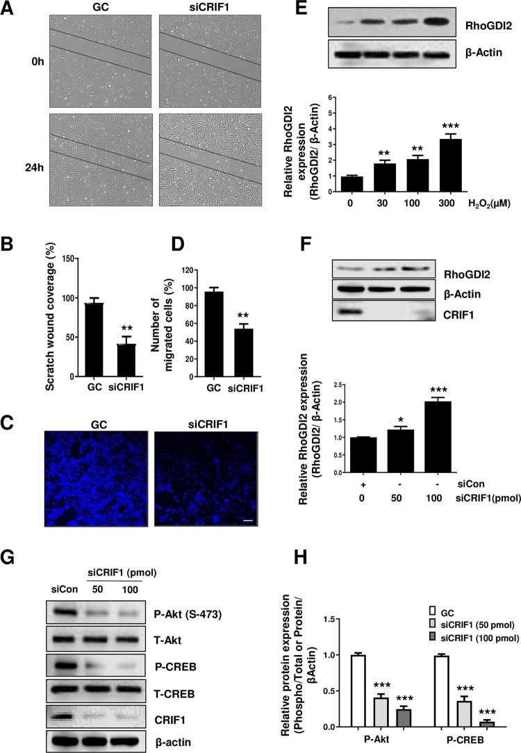
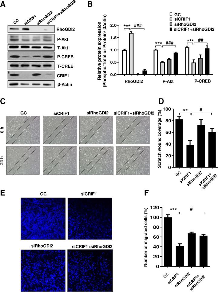
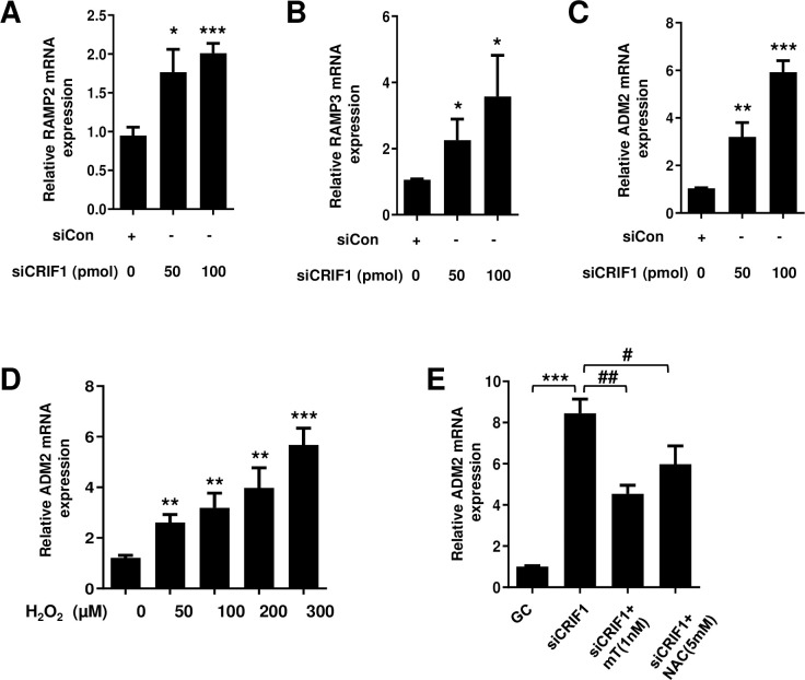
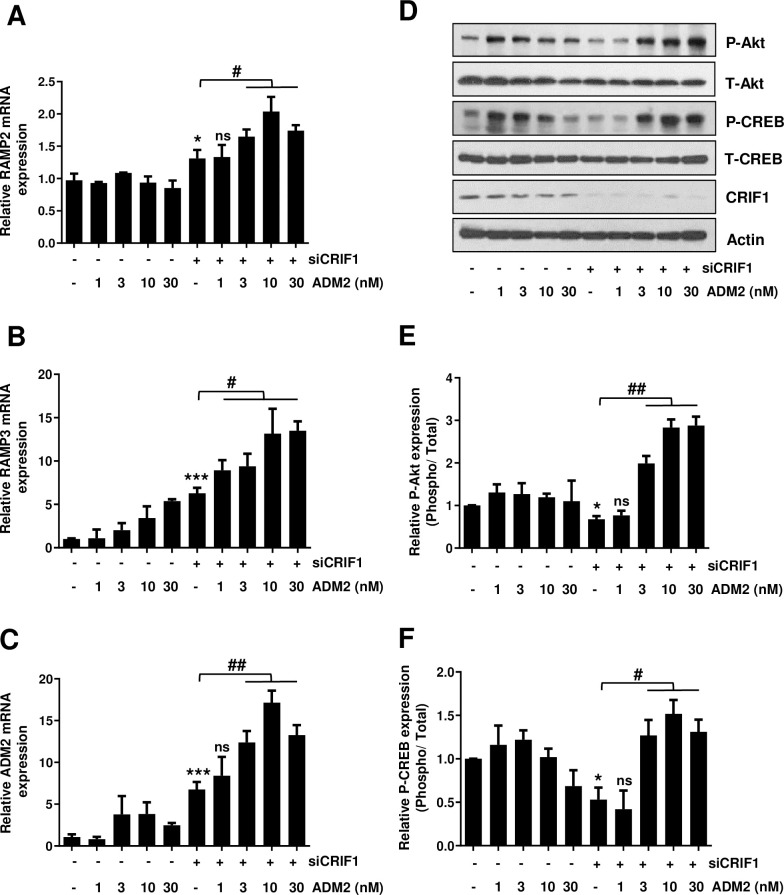
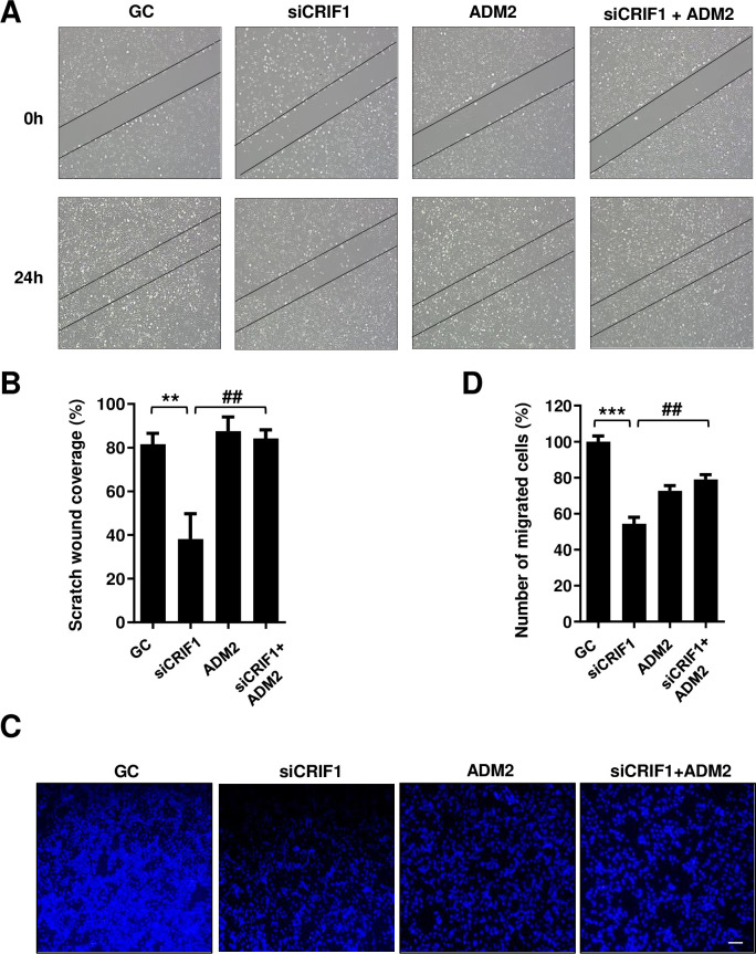
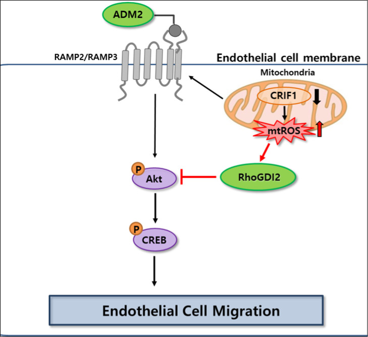
Rho GDP-dissociation inhibitor (RhoGDI), a downregulator of Rho family GTPases, prevents nucleotide exchange and membrane association. It is responsible for the activation of Rho GTPases, which regulate a variety of cellular processes, such as migration. Although RhoGDI2 has been identified as a tumor suppressor gene involved in cellular migration and invasion, little is known about its role in vascular endothelial cell (EC) migration. CR6-interacting factor 1 (CRIF1) is a CR6/GADD45-interacting protein with important mitochondrial functions and regulation of cell growth. We examined the expression of RhoGDI2 in CRIF1-deficient human umbilical vein endothelial cells (HUVECs) and its role in cell migration. Expression of RhoGDI2 was found to be considerably higher in CRIF1-deficient HUVECs along with suppression of cell migration. Moreover, the phosphorylation levels of Akt and CREB were decreased in CRIF1-silenced cells. The Akt-CREB signaling pathway was implicated in the changes in endothelial cell migration caused by CRIF1 downregulation. In addition to RhoGDI2, we identified another factor that promotes migration and invasion of ECs. Adrenomedullin2 (ADM2) is an autocrine/paracrine factor that regulates vascular tone and other vascular functions. Endogenous ADM2 levels were elevated in CRIF1-silenced HUVECs with no effect on cell migration. However, siRNA-mediated depletion of RhoGDI2 or exogenous ADM2 administration significantly restored cell migration via the Akt-CREB signaling pathway. In conclusion, RhoGDI2 and ADM2 play important roles in the migration of CRIF1-deficient endothelial cells.


Conflict of interest statement

The authors have declared that no competing interests exist.

Figures

Fig 1
Fig 1. Effect of CRIF1 deficiency on cell migration and RhoGDI2 expression in endothelial cells.
(A) HUVECs were cultured in six-well plates, transfected with control or CRIF1 siRNA (100 pmol), and incubated for 24 h. Next, the cells were wounded for 24 h. Images were obtained using a light microscope. (B) Quantification of wound closure was performed using ImageJ software. (C) HUVECs were transfected with control or CRIF1 siRNA (100 pmol) and transwell assay was conducted to determine cell migration. Scale bar 200 μm. (D) Quantification of the number of migrated cells was performed using ImageJ software. (E) Western blot analysis of RhoGDI2 in HUVECs treated with the indicated doses of H2O2 for 24 h. β-actin was used as an internal control. RhoGDI2 protein level quantified by densitometric analysis using ImageJ software is shown in the down panel. (F) Western blot analysis of RhoGDI2 after 48 h of dose-dependent CRIF1 siRNA transfection. β-actin was used as an internal control. RhoGDI2 protein level quantified by densitometric analysis using ImageJ software is shown in the down panel. (G) Western blot of phospho-Akt and phospho-CREB levels in HUVECs after 48 h of CRIF1 siRNA transfection. β-actin was used as an internal control. (H) Protein levels were quantified by densitometric analysis using ImageJ software. Data are means ± SD of three independent experiments. *P < 0.05, **P < 0.01 and *** P < 0.001 relative to the control (n = 3 per group).
Fig 2
Fig 2. RhoGDI2 knockdown in CRIF1-silenced HUVECs leads to activation of the Akt-CREB signaling pathway and restoration of endothelial cell migration.
(A) HUVECs were transfected with the control, CRIF1 siRNA, or co-transfected with CRIF1 and RhoGDI2 siRNA (100 pmol) for 48 h followed by western blot analysis of phospho-Akt and phospho-CREB levels. (B) Protein levels were quantified by densitometric analysis using ImageJ software. (C) HUVECs were transfected with the control, CRIF1 siRNA, or co-transfected with CRIF1 and RhoGDI2 siRNA (100 pmol) and incubated for 24 h. Next, the cells were wounded for 24 h. Images were obtained using a light microscope. (D) Quantification of wound closure was performed using ImageJ software. (E) HUVECs were transfected with the control, CRIF1 siRNA, or co-transfected with CRIF1 and RhoGDI2 siRNA (100 pmol) and transwell assay was conducted to determine cell migration. Scale bar 200 μm. (F) Quantification of the number of migrated cells was performed using ImageJ software. Data are means ± SD of three independent experiments. **P < 0.01 and *** P < 0.001 relative to the control, #P < 0.05, ##P < 0.01 and ### P < 0.001 relative to siCRIF1 (n = 3 per group).
Fig 3
Fig 3. Effect of CRIF1 deficiency on the mRNA levels of RAMPs and endogenous ADM2 in endothelial cells.
HUVECs were transfected with CRIF1 siRNA for 48 h. Next, the mRNA levels of (A) RAMP2 (B) RAMP3 and (C) ADM2 were determined by qPCR. (D) HUVECs were treated with the indicated doses of H2O2 for 24 h followed by the detection of ADM2 mRNA levels (E) HUVECs were pretreated with the antioxidants MitoTEMPO (1 nM) and NAC (5 mM) for 1 h followed by transfection with control or CRIF1 siRNA (100 pmol) for 48 h. mRNA levels of ADM2 were determined by qPCR. Data are means ± SD of three independent experiments. *P < 0.05, **P < 0.01 and *** P < 0.001 relative to the control, #P < 0.05 and ##P < 0.01 relative to siCRIF1 (n = 3 per group).
Fig 4
Fig 4. Effect of exogenous ADM2 on the Akt-CREB signaling pathway in CRIF1-silenced endothelial cells.
HUVECs were transfected with control or CRIF1 siRNA (100 pmol) followed by treatment with ADM2 for 48 h. Next, the mRNA levels of (A) RAMP2 (B) RAMP3 and (C) ADM2 were determined by qPCR. (D) Western blot analysis of phospho-Akt and phospho-CREB levels. (E and F) Protein levels were quantified by densitometric analysis using ImageJ software. Data are means ± SD of three independent experiments. *P < 0.05 and *** P < 0.001 relative to the control, #P < 0.05, ##P < 0.01 and ns = not significant relative to siCRIF1 (n = 3 per group).
Fig 5
Fig 5. Effect of exogenous ADM2 on the migration of CRIF1-silenced endothelial cells.
(A) HUVECs were transfected with control or CRIF1 siRNA (100 pmol) and treated with ADM2 for 24 h. Next, the cells were wounded for another 24 h. Images were obtained using a light microscope. (B) Quantification of wound closure was performed using ImageJ software. (C) HUVECs were transfected with control or CRIF1 siRNA (100 pmol), treated with ADM2 for 24 h.and transwell assay was conducted to determine cell migration. Scale bar 200 μm. (D) Quantification of the number of migrated cells was performed using ImageJ software. Data are means ± SD of three independent experiments. **P < 0.01 and *** P < 0.001 relative to the control, ##P < 0.01 relative to siCRIF1 (n = 3 per group).
Fig 6
Fig 6. Schematic model of the pathway involved in CRIF1 knockdown induced decrease in cell migration and the possible roles of RhoGDI2 and ADM2 in endothelial cell migration.

References

    1. Trepat X., Chen Z., and Jacobson K., Cell migration. Compr Physiol, 2012. 2(4): p. 2369–92. doi: 10.1002/cphy.c110012 - DOI - PMC - PubMed
    1. Nobes C.D. and Hall A., Rho, rac, and cdc42 GTPases regulate the assembly of multimolecular focal complexes associated with actin stress fibers, lamellipodia, and filopodia. Cell, 1995. 81(1): p. 53–62. doi: 10.1016/0092-8674(95)90370-4 - DOI - PubMed
    1. Scherle P., Behrens T., and Staudt L.M., Ly-GDI, a GDP-dissociation inhibitor of the RhoA GTP-binding protein, is expressed preferentially in lymphocytes. Proc Natl Acad Sci U S A, 1993. 90(16): p. 7568–72. doi: 10.1073/pnas.90.16.7568 - DOI - PMC - PubMed
    1. Gildea J.J., et al.., RhoGDI2 is an invasion and metastasis suppressor gene in human cancer. Cancer Res, 2002. 62(22): p. 6418–23. - PubMed
    1. Theodorescu D., et al.., Reduced expression of metastasis suppressor RhoGDI2 is associated with decreased survival for patients with bladder cancer. Clin Cancer Res, 2004. 10(11): p. 3800–6. doi: 10.1158/1078-0432.CCR-03-0653 - DOI - PubMed

Publication types

MeSH terms

Substances