. 2022 Jan:162:20-31.
doi: 10.1016/j.yjmcc.2021.08.008. Epub 2021 Aug 24.

Bottom-up proteomic analysis of human adult cardiac tissue and isolated cardiomyocytes

Affiliations

Bottom-up proteomic analysis of human adult cardiac tissue and isolated cardiomyocytes

Melinda Wojtkiewicz et al. J Mol Cell Cardiol. 2022 Jan.

Abstract

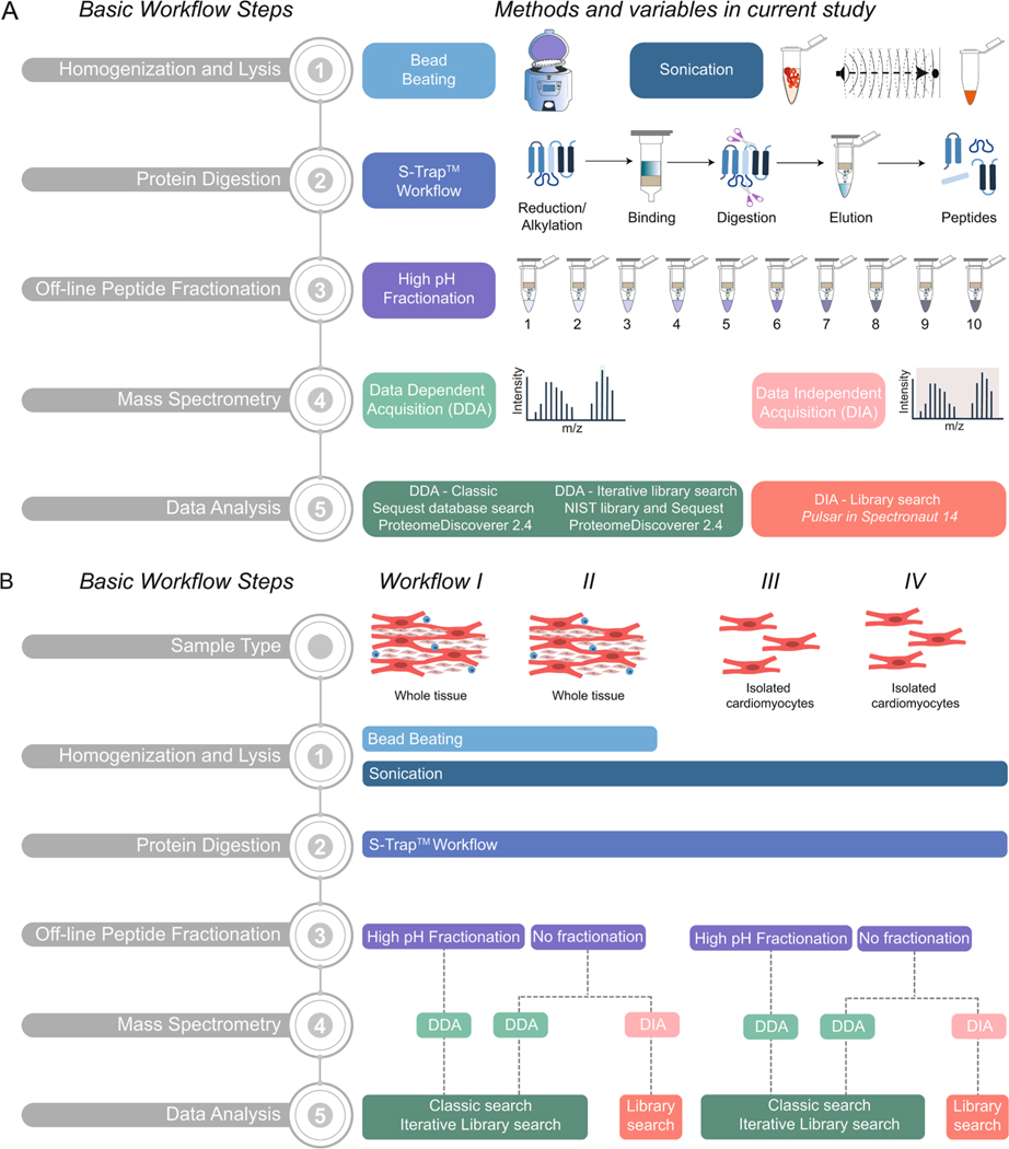
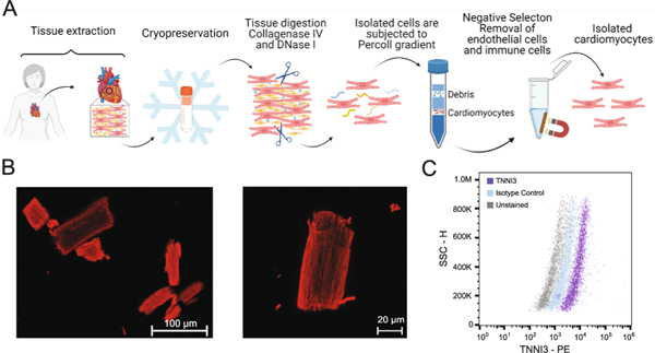
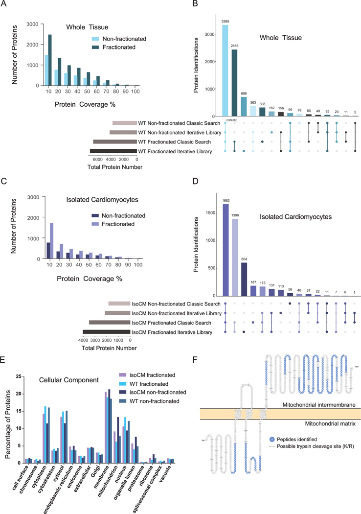
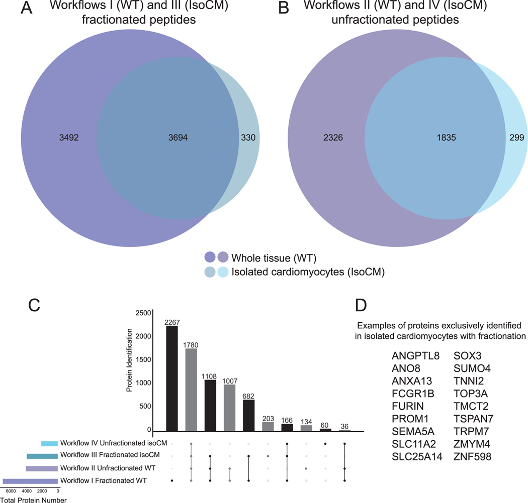
The heart is composed of multiple cell types, each with a specific function. Cell-type-specific approaches are necessary for defining the intricate molecular mechanisms underlying cardiac development, homeostasis, and pathology. While single-cell RNA-seq studies are beginning to define the chamber-specific cellular composition of the heart, our views of the proteome are more limited because most proteomics studies have utilized homogenized human cardiac tissue. To promote future cell-type specific analyses of the human heart, we describe the first method for cardiomyocyte isolation from cryopreserved human cardiac tissue followed by flow cytometry for purity assessment. We also describe a facile method for preparing isolated cardiomyocytes and whole cardiac tissue homogenate for bottom-up proteomic analyses. Prior experience in dissociating cardiac tissue or proteomics is not required to execute these methods. We compare different sample preparation workflows and analysis methods to demonstrate how these can impact the depth of proteome coverage achieved. We expect this how-to guide will serve as a starting point for investigators interested in general and cell-type-specific views of the cardiac proteome.

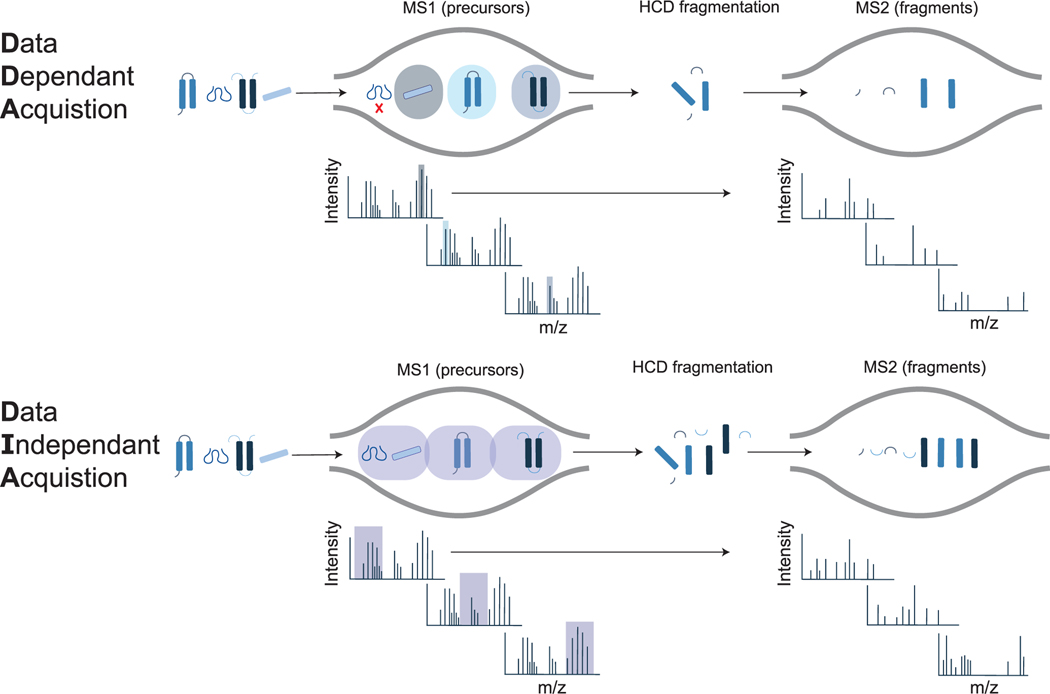
Keywords: Cardiomyocyte isolation; Data independent acquisition; Proteomics; S-trap.


Conflict of interest statement

Declaration of competing interest

R.L.G. is on the advisory board of ProtiFi, LLC and receives no compensation of any kind for this role.

Figures

Fig. 1.
Fig. 1.
Overview of bottom-up proteomics methodologies for processing human cardiac tissue and cardiomyocytes. A) Schematic representation of five basic steps in a bottom-up proteomics workflow: (1) Homogenization and Lysis, (2) Protein Digestion, (3) Off-line Peptide Fractionation, (4) Mass Spectrometry, and (5) Data Analysis. B) Schematic representation of four workflows applied for the proteomic analysis of human cardiac tissue or isolated cardiomyocytes in the current study.
Fig. 2.
Fig. 2.
Cardiomyocyte isolation and purity assessment. A) Schematic overview of cardiomyocyte isolation workflow using cryopreserved human adult cardiac tissue. B) Representative confocal immunofluorescence images of isolated cardiomyocytes stained for cardiac actin (red). Magnification 20× (left) and 63× (right); Scale bar, 100 μm (left), 20 μm (right) C) Flow cytometry assessment of isolated cells for TNNI3 positivity. Cells gated on fluorescence intensity versus side scatter (SSC-H) result in a >80% anti-TNNI3 PE positive population as compared to isotype control. Panel A was generated with Biorender. (For interpretation of the references to colour in this figure legend, the reader is referred to the web version of this article.)
Fig. 3.
Fig. 3.
Overview of whole tissue and isolated cardiomyocyte proteome. A) Bar chart displaying number of proteins identified at different percentages of coverage in workflows I and II. B) UpSet plot visualizing the intersections of identified proteins from workflows I and II in DDA mode, processed with a classic search and an iterative spectral library search. C) Bar chart displaying number of proteins identified at different percentages of coverage in both workflow III and IV in DDA mode. D) UpSet plot visualizing the intersections of identified proteins from workflow III and IV in DDA mode, processed with a classic search and an iterative spectral library search. E) Percentage of proteins annotated among various cellular components based on Gene Ontology for each workflow. F) Protein sequence showing 34% coverage of mitochondrial potassium channel ATP-binding subunit (ABCB8). For each sample type, three biological replicates (donors) were each analyzed by three technical replicates by MS. UpSet plots were generated using UpSetR [34]. Note: Upset plots graph the intersections of various elements, similar to a Venn diagram. However, unlike a Venn diagram, an upset plot is not limited by a certain number of elements and provides an easier way to interpret the common and uncommon identifications in proteomic datasets.
Fig. 4.
Fig. 4.
Deeper view of the proteome is achieved when peptide fractionation is combined with cardiomyocyte isolation. A and B) Venn diagrams displaying total number of proteins identified in apex for each workflow by DDA searched with the iterative spectral library strategy. C) UpSet Plot (generated by UpSetR [34]) showing overlap of proteins identified in unfractionated and fractionated apex whole tissue and isolated cardiomyocytes from apex. D) Examples from among the 203 proteins identified exclusively in isolated cardiomyocytes with fractionation.
Fig. 5.
Fig. 5.
Overview of DDA and DIA acquisition methods. In DDA mode, the instrument selects precursors for fragmentation one at a time, which can lead to missing features. In DIA mode, all precursors within a designated m/z window are fragmented sequentially and repeatedly throughout the acquisition, leading to fewer missing features, but complex MS2 spectra.
Fig. 6.
Fig. 6.
Overview of differentially abundant proteins in homogenized cardiac tissue and isolated cardiomyocytes using workflow II and IV in DIA mode. Volcano plot displaying differentially abundant proteins between A) left ventricular and right ventricular tissue and B) isolated cardiomyocytes from left and right ventricles. Horizontal bar represents a significance level of Qvalue ≤0.05. Vertical bars represent a significance level of log2 fold change ≥0.58 (right) and ≤ − 0.58 (left). Log2 intensities measurements reveal proteins that were higher in abundance in C) left ventricular cardiomyocytes (ANKRD2, ABAT, CPLV, ATP5MC1) and D) tissue. E) ATP5MC1 was not significantly more abundant when comparing left and right ventricular tissue. Log2 intensities measurements reveal proteins that were higher in abundance in F) right ventricular cardiomyocytes (CHDH, MRPL20, HRNR, CALML5) and G) tissue. H) MRPL20 was not significantly more abundant when comparing left and right ventricular tissue. Purple asterisk indicates proteins not detected in the quantitative analysis of whole tissue. Green asterisk indicates proteins that were quantified by 1 peptide. All other proteins were quantified by 2 or more peptides. For each sample type, three biological replicates (donors) were analyzed by three technical replicates by MS. (pairwise t-test; *p < 0.05, **p < 0.01). (For interpretation of the references to colour in this figure legend, the reader is referred to the web version of this article.)

References

    1. Fountoulaki K, Cardiothoracic Intensive Care Unit, Onassis Cardiac Surgery Centre, Athens, Greece, N. Dagres, Second University Department of Cardiology, Attikon General Hospial, University of Athens, Athens, Greece, Cellular communications in the heart, Card. Fail. Rev 1 (2015) 64, 10.15420/cfr.2015.1.2.64. - DOI - PMC - PubMed
    1. Martins-Marques T, Hausenloy DJ, Sluijter JPG, Leybaert L, Girao H, Intercellular communication in the heart: therapeutic opportunities for cardiac ischemia, Trends Mol. Med 27 (2021) 248–262, 10.1016/j.molmed.2020.10.002. - DOI - PubMed
    1. Asp M, Giacomello S, Larsson L, Wu C, Fürth D, Qian X, Wärdell E, Custodio J, Reimegård J, Salmén F, Österholm C, Ståhl PL, Sundström E, Åkesson E, Bergmann O, Bienko M, Månsson-Broberg A, Nilsson M, Sylvén C, Lundeberg J, A spatiotemporal organ-wide gene expression and cell atlas of the developing human heart, Cell 179 (2019) 1647–1660.e19, 10.1016/j.cell.2019.11.025. - DOI - PubMed
    1. Tucker Nathan R., Mark Chaffin, Stephen J. Fleming, Amelia W. Hall, Victoria A. Parsons, Kenneth C. Bedi, Amer-Denis Akkad, Caroline N. Herndon, Alessandro Arduini, Irinna Papangeli, Carolina Roselli, François Aguet, Hoan Choi Seung, Kristin G. Ardlie, Mehrtash Babadi, Kenneth B. Margulies, Christian M. Stegmann, Patrick T. Ellinor, Transcriptional and cellular diversity of the human heart, Circulation 142 (2020) 466–482, 10.1161/CIRCULATIONAHA.119.045401. - DOI - PMC - PubMed
    1. Litviñuková M, Talavera-López C, Maatz H, Reichart D, Worth CL, Lindberg EL, Kanda M, Polanski K, Heinig M, Lee M, Nadelmann ER, Roberts K, Tuck L, Fasouli ES, DeLaughter DM, McDonough B, Wakimoto H, Gorham JL, Samari S, Mahbubani KT, Saeb-Parsy K, Patone G, Boyle JJ, Zhang H, Zhang H, Viveiros A, Oudit GY, Bayraktar OA, Seidman JG, Seidman CE, Noseda M, Hubner N, Teichmann SA, Cells of the adult human heart, Nature 588 (2020) 466–472, 10.1038/s41586-020-2797-4. - DOI - PMC - PubMed

Publication types