Review
. 2021 Jul 31;11(8):2271.
doi: 10.3390/ani11082271.

Spatial Cognition in Teleost Fish: Strategies and Mechanisms

Affiliations
Review

Spatial Cognition in Teleost Fish: Strategies and Mechanisms

Fernando Rodríguez et al. Animals (Basel). .

Abstract

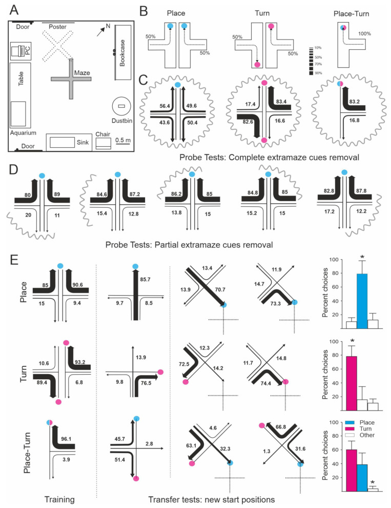
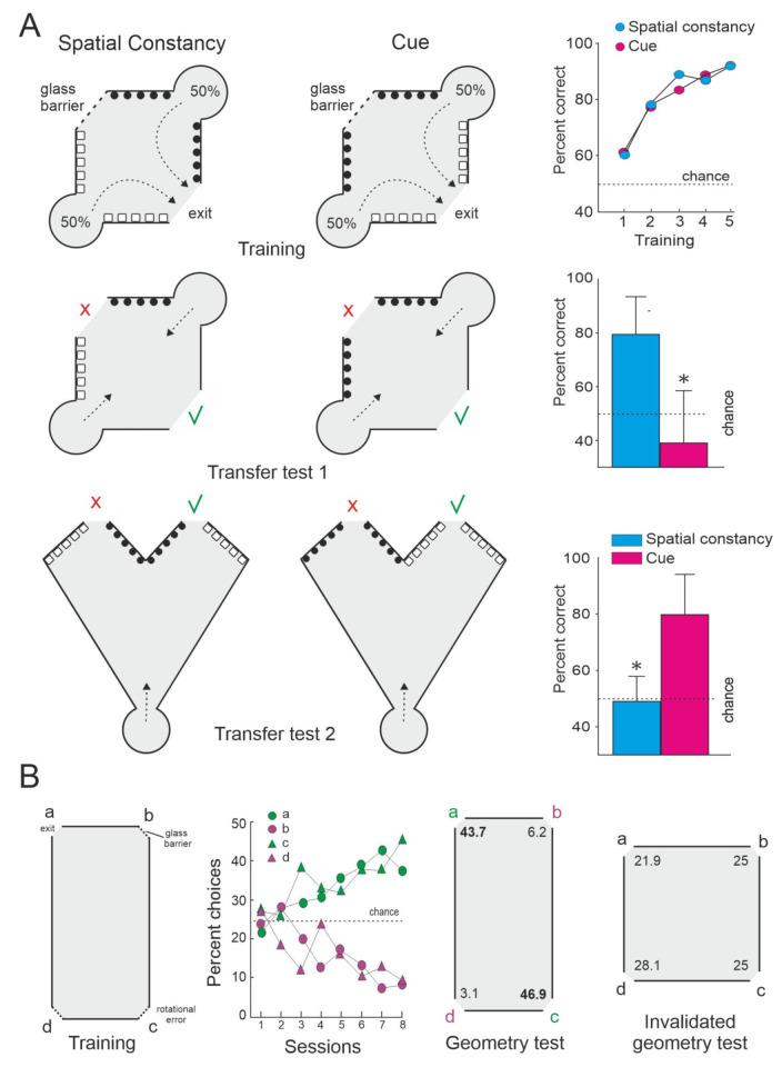
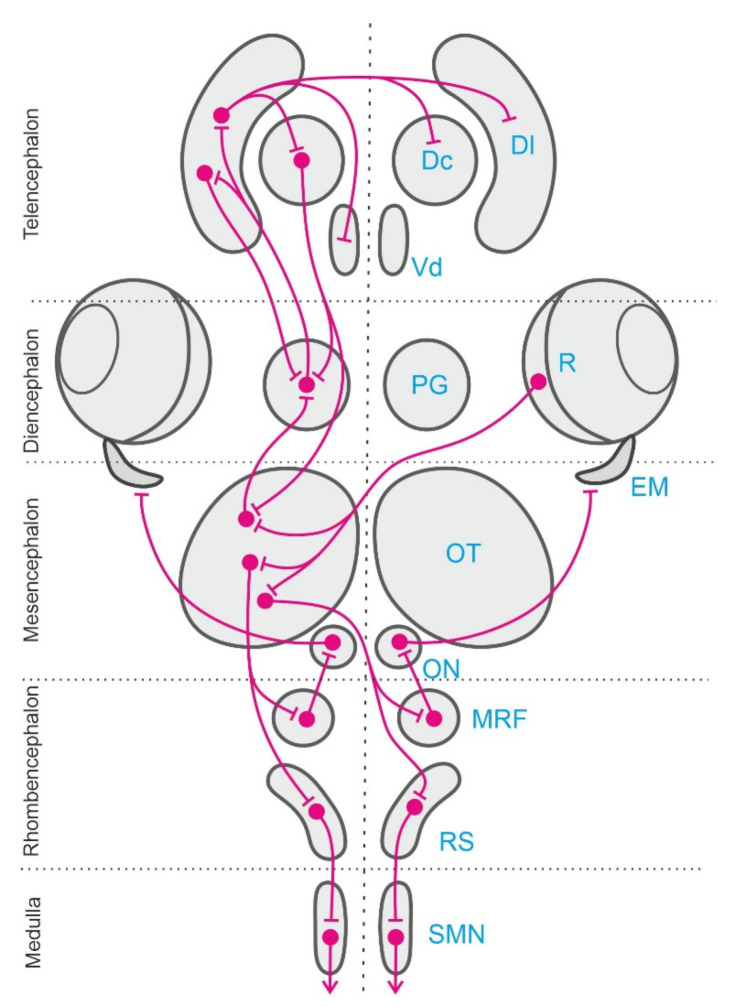
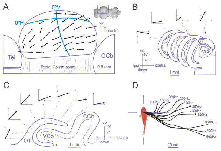
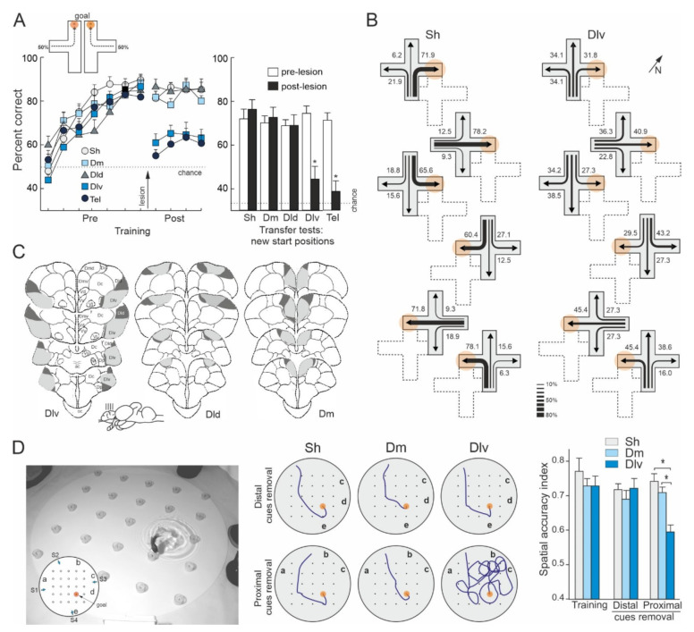
Teleost fish have been traditionally considered primitive vertebrates compared to mammals and birds in regard to brain complexity and behavioral functions. However, an increasing amount of evidence suggests that teleosts show advanced cognitive capabilities including spatial navigation skills that parallel those of land vertebrates. Teleost fish rely on a multiplicity of sensory cues and can use a variety of spatial strategies for navigation, ranging from relatively simple body-centered orientation responses to allocentric or "external world-centered" navigation, likely based on map-like relational memory representations of the environment. These distinct spatial strategies are based on separate brain mechanisms. For example, a crucial brain center for egocentric orientation in teleost fish is the optic tectum, which can be considered an essential hub in a wider brain network responsible for the generation of egocentrically referenced actions in space. In contrast, other brain centers, such as the dorsolateral telencephalic pallium of teleost fish, considered homologue to the hippocampal pallium of land vertebrates, seem to be crucial for allocentric navigation based on map-like spatial memory. Such hypothetical relational memory representations endow fish's spatial behavior with considerable navigational flexibility, allowing them, for example, to perform shortcuts and detours.

Keywords: hippocampal pallium; optic tectum; spatial navigation; spatial strategies; telencephalon; teleost fish; vertebrate brain evolution.


Conflict of interest statement

The authors declare no conflict of interest.

Figures

Figure 1
Figure 1
Spatial navigation strategies used by goldfish to solve different procedures in a four-arm maze. (A) Experimental room showing the maze in its training position (solid line) and in its rotated and displaced position used in the transfer tests (dotted line), and the extramaze cues. (B) Training procedures. Arrows show the most effective path to reach the goal. Place and turn procedures used two different start positions randomly assigned across trials (50% each). The colored circle marks the goal location in each procedure. (C) Percentage of choices in the probe test in which all the extramaze cues were occluded by means of curtains. The numbers and the relative thickness of the arrows denote the percentage of times that a particular choice was made. (D) Percentage of choices by the animals in the place procedure in the probe tests in which only a part of the extramaze cues were occluded. (E) Trajectories chosen by the animals in the different groups during training and transfer trials in which new start positions were employed. In one type of transfer tests (left) the maze remained in its usual position, in the other type (right), the maze was displaced in the room in such a way that the end of one arm was located in the same place where the fish were rewarded during training trials. The dashed lines indicate the original position of the maze during training. The blue circles mark the goal place for animals in the place and the place-turn procedures. The red circles mark the goal for the turn group during training and the arm corresponding with an egocentric (turn) strategy for both the turn and the place-turn procedures. The histograms on the right show the accumulated mean percentage of choices during the transfer tests. Asterisks denote significant differences. Modified from [17].
Figure 2
Figure 2
Relational map-like spatial representations in small stimulus-controlled mazes. (A) Two group of goldfish were trained to exit from an enclosure in a spatial constancy task which requires the use of allocentric (relational) strategies or in a cued version of the same task. The access from the start compartments, the distribution of the experimental visual cues (black and white symbols), the position of the glass barrier, and the location of the goal (exit) are shown for both training procedures. The numbers indicate the percentage of trials initiated from each start compartment. The arrows show the most efficient trajectories to the goal. Note that in the transfer tests the deletion of the local cues directly associated with the goal (Transfer Test 1) did not alter the performance in the relational task (spatial constancy), however the alteration of the global layout of the experimental setup (Transfer test 2) disrupted performance, even though the relationships between the local cues and the goal remained unaltered in the transfer tests. The green check marks indicate the door corresponding to the goal during training conditions. The figures on the right show the percentage of correct responses during training and transfer tests. Asterisks denote significant differences. Modified from [16]. (B) Encoding of geometrical spatial information by goldfish. Fish were trained to find the exit door (goal) placed in a corner (a) of a rectangular environment on the basis of the geometrical information provided by the apparatus. The arena had three identical, blocked openings (glass barriers) in the other three corners (b–d). Note that because of the geometric properties of the apparatus, the correct corner was indistinguishable from the diagonally opposite (180°) corner (rotational error). The percentage of choices for the four corners during training is shown. Two different probe trials were carried out in which the glass barriers were not used, so that fish could exit freely through any door. For the invalidated geometry test, a new apparatus that modified the geometric properties of the experimental enclosure was used. Numbers in the diagrams indicate the percentage of choices to each door during the tests. Modified from [87].
Figure 3
Figure 3
Schematic representation of the extended neural network involved in the egocentric sensorimotor transformations and egocentric-to-allocentric spatial reference framework conversion described in the text. Only the left half of the bilateral network is presented here. The right retina (R) sends visual information to the contralateral optic tectum (OT). Visual and other sensory modalities converge into the OT and in other stages of this neural network, where multisensory integration takes place. The multisensory information is represented in the OT in a body-centered map. Sensorimotor transformations leading to the generation of the egocentric orienting responses are performed at early processing stages in the OT, but also in parallel and sequentially in other nodes of the network. The tectal motor commands, encoded in egocentric coordinates, are conveyed to the neural circuits in the mesencephalic reticular formation (MRF) that organize the saccadic motor programs. These signals are submitted to further transformations into the OT-MRF interface to adapt to the specific requirements of the ocular motor plant, and in turn they finally activate the extraocular motoneurons in the oculomotor nuclei (ON) to produce orienting eye-movements (EM, extraocular muscles). Tectal efferences also activate reticulospinal (RS) assemblies, which in turn recruit spinal motor networks (SMN) to produce orienting (ipsilateral descending pathway) or avoidance (contralateral descending pathway) responses. In regard to the ascending (prosencephalic) tectal projections, the OT send massive efferents to the preglomerular complex (PG), the main diencephalic sensory relay station of teleost fish, which in turn project into the intricate telencephalic neural networks in which the egocentric-to-allocentric reference framework transformations are thought to take place. The main visual information recipient pallial region is the dorsolateral area (Dl), which has demonstrated to be a crucial center for allocentric navigation, with other sensory modalities reaching separate pallial targets. The telencephalic pallium sends back outputs to the PG, the OT, and other descending motor networks through the dorsocentral (Dc) pallial area and through other telencephalic subpallial structures, as the ventrodorsal nucleus (Vd) of the telencephalon, an area likely homologous to the tetrapod’s basal ganglia, which finally modulates behavioral responses.
Figure 4
Figure 4
The optic tectum of teleost fish is a crucial brain center for the generation of egocentric orientation responses. Electrical microstimulation in the optic tectum of goldfish elicits coordinated eye (AC) and body (D) movements. (A) Vectorial representation (black arrows) of the evoked eye movements after focal electrical stimulation in the optic tectum, showing the amplitude and direction of the saccades. Note that the amplitude and direction of eye movement vectors depend on the stimulation site within the tectum. The retinotopic vertical and horizontal axis are superimposed (blue lines). (B) Variation of the stimulation site in the medial–lateral axis produces an increase in the vertical component. (C) Variation in the stimulation sites across the rostro–caudal axis produced a systematic change in the amplitude of the horizontal component of the saccade. (D) The direction and amplitude of the orienting responses in free-swimming fish depend on both the tectal stimulation site and the stimulus parameters. The insert in (A) shows a dorsal view of the goldfish brain and the square marks the magnified area. CCb: corpus cerebellum; OT: optic tectum; Tel: telencephalon; VCb: valvula cerebellum; ipsi and contra: ipsiversive and contraversive direction of evoked eye saccade, respectively. Modified from [131,133].
Figure 5
Figure 5
Dorsolateral telencephalon lesions in teleost fish produce significant deficits in spatial navigation tasks that require the use of map-like strategies. (A) Effects of different pallial lesions on the learning of a place task in a plus maze (see Figure 1). The curves show the mean percentage of correct choices during pre- and post-lesion training sessions. The histograms show the percentage of correct choices during the transfer trials in which new start positions were employed. Asterisks denote significant differences. (B) Trajectories chosen by sham-operated (Sh) and dorsolateral telencephalon-lesioned (Dlv) goldfish during the transfer trials conducted after surgery, in which the maze was displaced, and new start positions were employed. The numbers and the relative thickness of the arrows denote the percentage of times that a particular choice was made. The position of the maze during training trials is shown by dotted lines. Note that the Sh goldfish consistently chose the route leading to the place where they were rewarded during the training trials (orange circle). In contrast, the random distribution of the choices by Dlv-lesioned animals revealed a profound spatial deficit. (C) Schematic transversal drawings of the telencephalon of goldfish showing the largest (dark grey) and the smallest (light grey) extensions of the different pallial lesions. Dm: dorsomedial telencephalon-lesioned group; tel: telencephalon ablated group. Modified from [157]. (D) Effects of dorsomedial (Dm) and dorsolateral (Dl) telencephalon lesions in the use of allocentric strategies to locate a goal in a hole-board homologue task. At the right is shown a photograph of the experimental apparatus and the training procedure. In the insert is shown the goal (baited feeder, red circle), the position of the cues (letters a–e), and the four different start positions used during training (S1–S4). The diagrams on the right show the searching trajectories of a representative fish of each group on the distal and proximal cue-removal tests. Note the spatial deficit of Dlv goldfish when the cues in the vicinity of the goal were removed. The histogram shows the mean spatial accuracy index (values relative to distance to the goal) in training and in the probe tests in which the distal (a,b) or the proximal (d,e) cues to the goal were removed. Asterisks denote significant differences. Modified from [182].

References

    1. Dodson S. The ecological role of chemical stimuli for the zooplankton: Predator-avoidance behavior in Daphnia. Limnol. Oceanogr. 1988;33:1431–1439. doi: 10.4319/lo.1988.33.6_part_2.1431. - DOI
    1. Quinn T.P. Fishes. In: Papi F., editor. Animal Homing. Chapman & Hall; London, UK: 1992. pp. 145–211.
    1. Hallacher L.E. Relocation of original territories by displaced black-and-yellow rockfish, Sebastes chrysomelas, from Carmel Bay, California. Calif. Fish Game. 1984;7:158–162.
    1. Kroon F.J., de Graaf M., Liley N.R. Social organisation and competition for refuges and nest sites in Coryphopterus nicholsii (Gobiidae), a temperature protogynous reef fish. Environ. Biol. Fishes. 2000;57:401–411. doi: 10.1023/A:1007600728267. - DOI
    1. Matthews K.R. An experimental study of the habitat preferences and movement patterns of copper, quillback, and brown rockfishes (Sebastes spp.) Environ. Biol. Fishes. 1990;29:161–178. doi: 10.1007/BF00002217. - DOI

LinkOut - more resources