Review
. 2021 Aug 4;13(16):3921.
doi: 10.3390/cancers13163921.

Vitreoretinal Lymphoma

Affiliations
Review

Vitreoretinal Lymphoma

Bianka Sobolewska et al. Cancers (Basel). .

Abstract

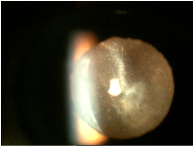
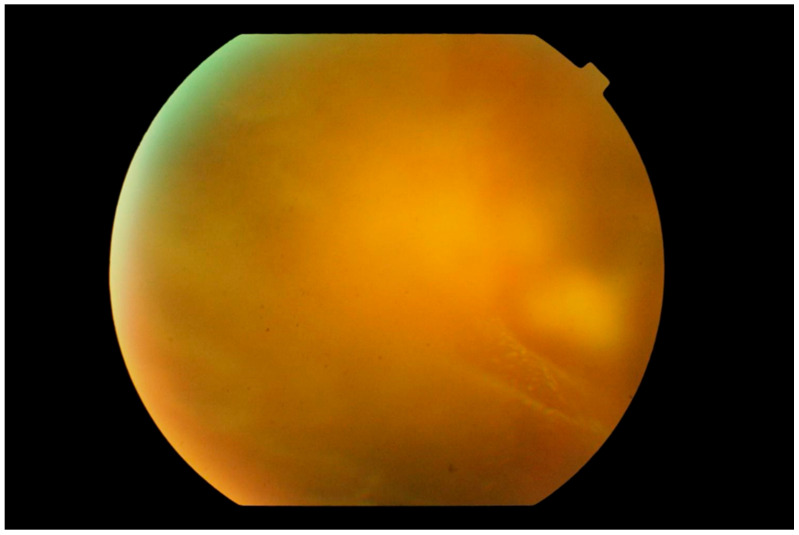
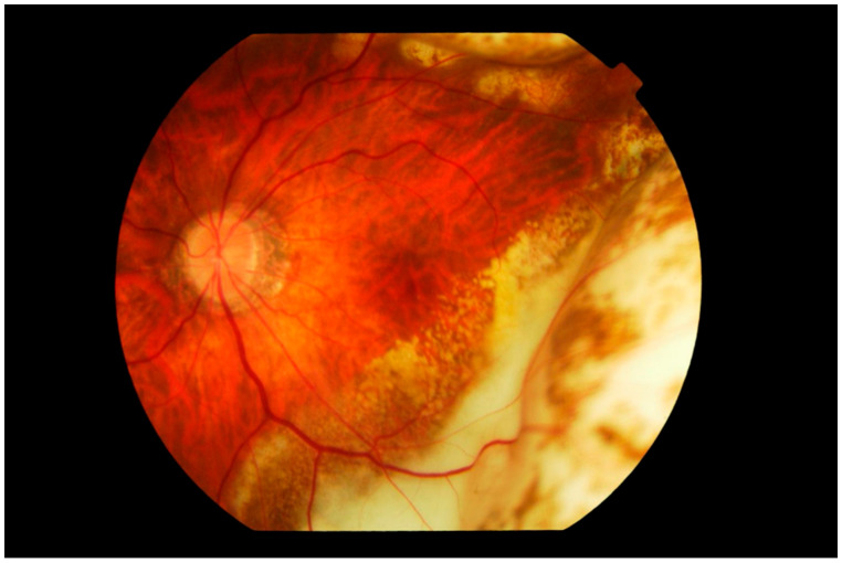
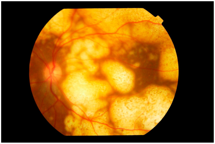
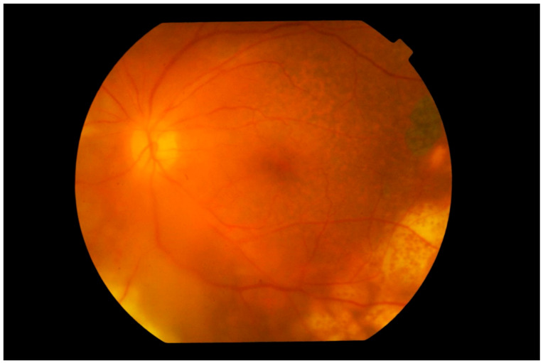
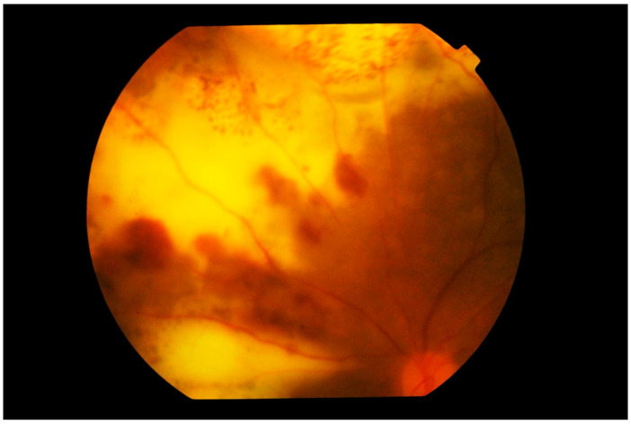
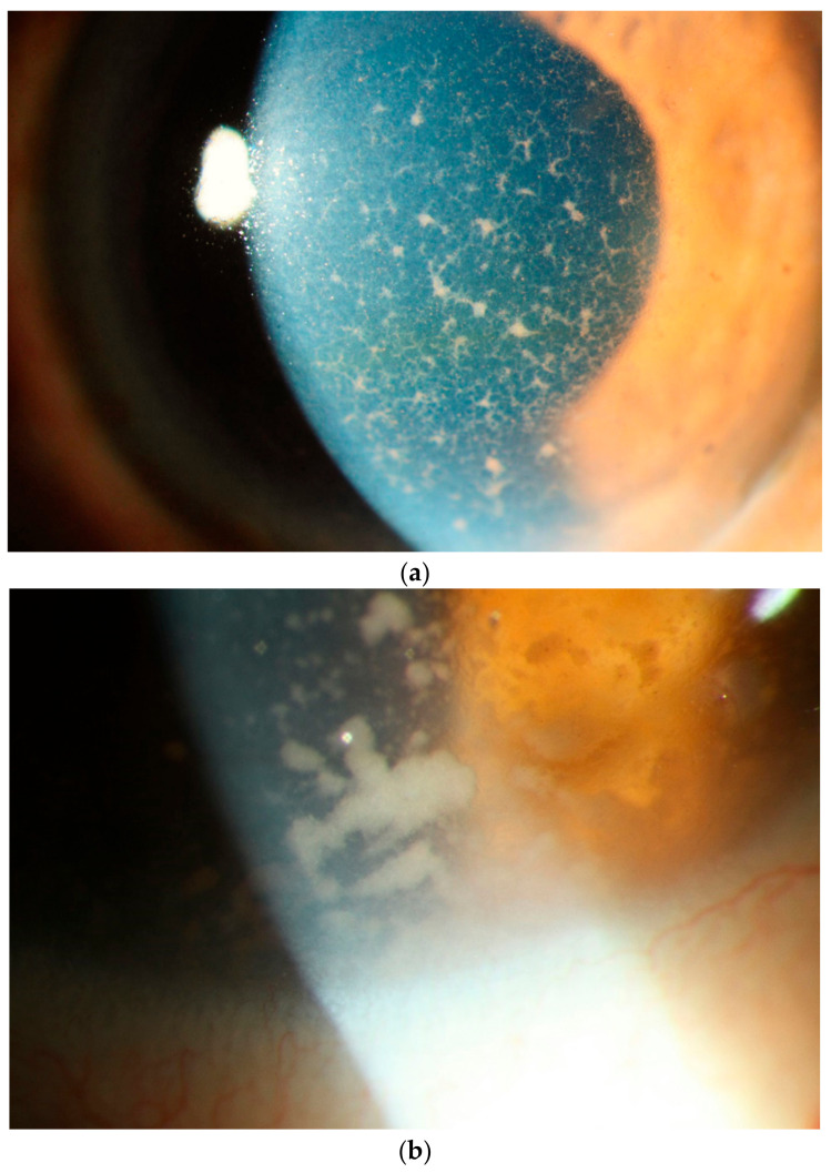
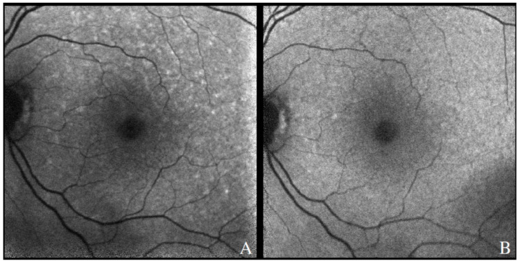
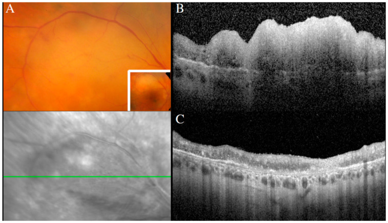
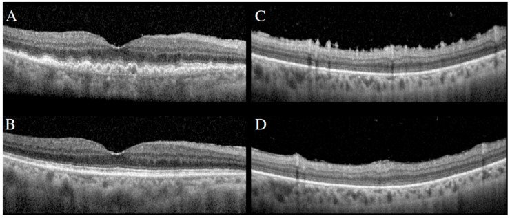
Vitreoretinal lymphoma (VRL) is a rare variant of primary central nervous system lymphoma (PCNSL), mostly of diffuse large B cell lymphoma, which affects the retina and/or the vitreous with or without optic nerve involvement. The disease course is aggressive. Up to 90% of the patients develop central nervous system lymphoma within one year. The diagnosis of VRL is challenging due to nonspecific chronic and relapsing uveitis and is made by anterior chamber tab or vitreous aspirate biopsy. There is no established treatment protocol for VRL patients with bilateral involvement without CNS involvement. There are suggestions to use only intravitreal chemotherapy with methotrexate and/or rituximab. Alternatively, systemic high-dose MTX treatment or external beam radiotherapy is used. Further studies are needed to prove and confirm the prophylactic systemic therapy in preventing CNS involvement in limited VRL.

Keywords: CNS lymphoma; IL-10/IL-6 ratio; MYD88; methotrexate; vitreoretinal lymphoma.


Conflict of interest statement

The authors declare no conflict of interest.

Figures

Figure 1
Figure 1
Slit lamp photograph of the retrolental region demonstrating sheets of numerous white cells of varied sizes infiltrating the vitreous. Most of these lymphoma cells tend to be larger than what is seen in vitritis.
Figure 2
Figure 2
Photograph of the peripheral fundus showing the “aurora borealis” sign caused by lymphoma cell infiltration along the vitreous fibrils creating streaks of opacity. In the background, a multinodular yellowish mass is just visible.
Figure 3
Figure 3
Fundus photograph of the left eye showing subretinal infiltration by lymphoma cells forming multiple large yellowish cream nodular masses distributed circumferentially with patchy pigmentation, giving a characteristic “leopard spot” appearance. The inferior temporal fundus shows the recent development of a wide span of subretinal infiltrates, contiguous with the longer standing peripheral mass. The edge of the lesion advancing towards the fovea consists of multiple small cream-colored spots. The vitreous is clear.
Figure 4
Figure 4
Fundus photograph of the left eye showing extensive involvement of the entire fundus by multiple nodular subretinal lymphoma masses of varying sizes. The larger peripheral lesions are formed by the merging of small lesions and have pigmentary change, giving a “leopard spot” appearance. The multiple yellow creamy lesions superior temporal to the disc which are smaller and more recent are also subretinal in location.
Figure 5
Figure 5
Photograph of the left fundus showing multiple yellowish nodular subretinal lesions with “leopard spot” pigmentation inferior temporally. Temporally to the fovea and in the superior temporal fundus, pigmentary mottling of the retina representing previous spontaneously resolved vitreoretinal lymphoma lesions is clearly seen. The vitreous is hazy but the disc appears normal.
Figure 6
Figure 6
Photograph of the left superior nasal fundus showing large confluent yellowish subretinal infiltration by lymphoma cells. Large blotches of retinal hemorrhage are also observed, creating a picture of deep retinal necrosis. Note that the vitreous is clear and the disc is not swollen.
Figure 7
Figure 7
Slit lamp photograph showing (a) diffusely distributed keratic precipitates (KP) on the corneal endothelium. These KPs are a mixture of small and fine or infiltrative KP intermixed with some granulomatous KP. Many of these granulomatous KP have fibrillar extensions, taking on a comet-like appearance, typical of vitreoretinal lymphoma. These faintly pigmented KP may be mistaken for the KP of viral anterior uveitis. (b) Occasionally one can find even larger tumor cell collections on the endothelium.
Figure 8
Figure 8
Fundus autofluorescence demonstrating hyperautofluorescent spots in the posterior pole of a patient with recurrence of PVRL (A). There was resolution after therapy involving systemic chemotherapy, with few residual hypoautofluorescent spots along the superior arcade (B).
Figure 9
Figure 9
Creamy retinal lesion on a fundus photograph (A), corresponding to a large inner retinal infiltrate on a SD-OCT scan (B). Regression of the retinal lesion after intravitreal methotrexate, with residual retinal atrophy (C).
Figure 10
Figure 10
SD-OCT demonstrating large sub-RPE infiltrates (A) and preretinal/inner retinal spike-like lesions (C). Following intravitreal methotrexate, resolution of sub-RPE infiltrates (B) and spike-like lesions (D).

References

    1. Cao X., Shen D., Callanan D.G., Mochizuki M., Chan C.-C. Diagnosis of systemic metastatic retinal lymphoma. Acta Ophthalmol. 2011;89:e149–e154. doi: 10.1111/j.1755-3768.2009.01797.x. - DOI - PMC - PubMed
    1. Tiu Teo H.M., Çiftçi S., Elner V.M., Demirci H. Systemic anti-CD20 (rituximab) as primary treatment for symptomatic primary uveal lymphoma. Am. J. Ophthalmol. Case Rep. 2019;15:100484. doi: 10.1016/j.ajoc.2019.100484. - DOI - PMC - PubMed
    1. Salomão D.R., Pulido J.S., Johnston P.B., Canal-Fontcuberta I., Feldman A. Vitreoretinal Presentation of Secondary Large B-Cell Lymphoma in Patients with Systemic Lymphoma. JAMA Ophthalmol. 2013;131:1151–1158. doi: 10.1001/jamaophthalmol.2013.334. - DOI - PubMed
    1. Jahnke K., Korfel A., Komm J., Bechrakis N.E., Stein H., Thiel E., Coupland S.E. Intraocular lymphoma 2000–2005: Results of a retrospective multicentre trial. Graefe’s Arch. Clin. Exp. Ophthalmol. 2006;244:663–669. doi: 10.1007/s00417-005-0138-9. - DOI - PubMed
    1. Fonkem E., Lok E., Robison D., Gautam S., Wong E.T. The natural history of intravascular lymphomatosis. Cancer Med. 2014;3:1010–1024. doi: 10.1002/cam4.269. - DOI - PMC - PubMed

LinkOut - more resources