. 2021 Aug 14;13(16):4095.
doi: 10.3390/cancers13164095.

Smart Modification on Magnetic Nanoparticles Dramatically Enhances Their Therapeutic Properties

Affiliations

Smart Modification on Magnetic Nanoparticles Dramatically Enhances Their Therapeutic Properties

Nuria Lafuente-Gómez et al. Cancers (Basel). .

Abstract

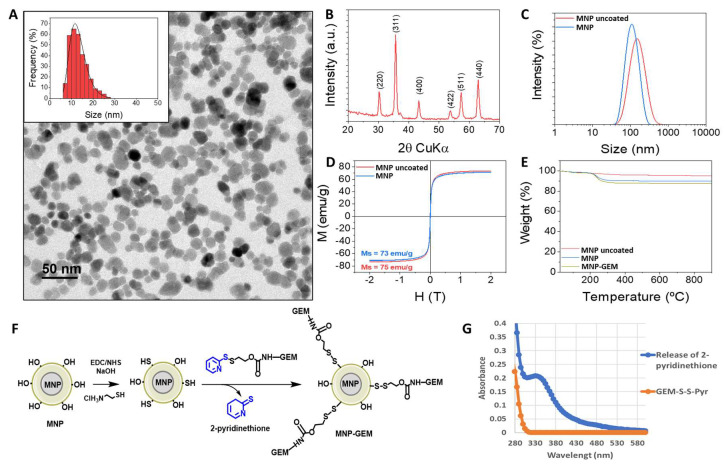
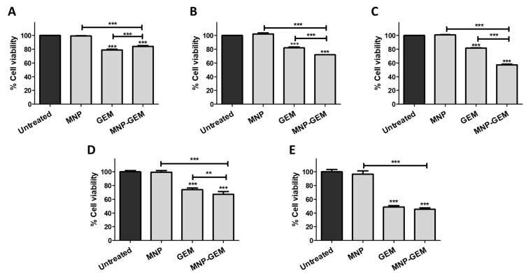
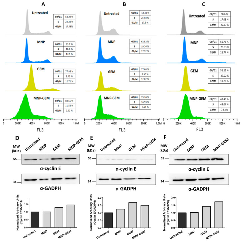
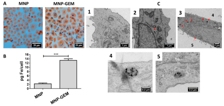
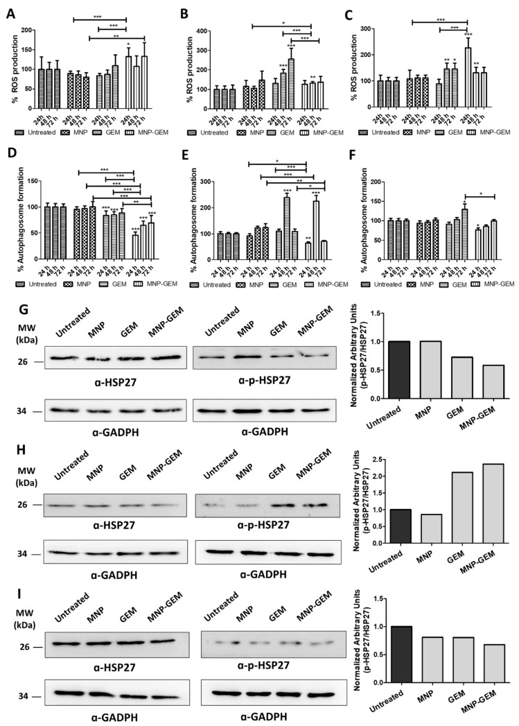
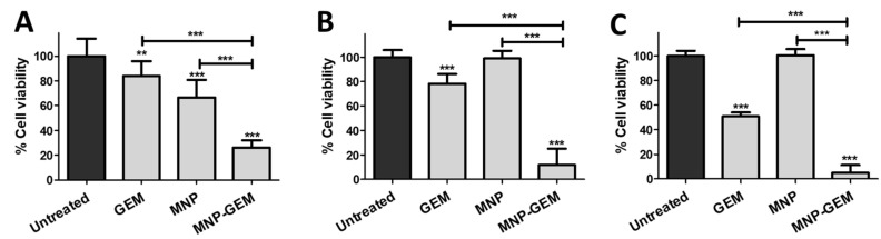
Magnetic nanoparticles (MNP) are employed as nanocarriers and in magnetic hyperthermia (MH) for the treatment of cancers. Herein, a smart drug delivery system composed of MNP functionalized with the cytotoxic drug gemcitabine (MNP-GEM) has been thoroughly evaluated. The linker employed is based on a disulfide bond and allows the controlled release of GEM under a highly reducing environment, which is frequently present in the cytoplasm of tumor cells. The stability, MH, and the interaction with plasma proteins of the nanoparticles are evaluated, highlighting their great potential for biological applications. Their cytotoxicity is assessed in three pancreatic cancer cell lines with different sensitivity to GEM, including the generation of reactive oxygen species (ROS), the effects on the cell cycle, and the mechanisms of cell death involved. Remarkably, the proposed nanocarrier is better internalized than unmodified nanoparticles, and it is particularly effective in PANC-1 cells, resistant to GEM, but not in non-tumoral keratinocytes. Additionally, its combination with MH produces a synergistic cytotoxic effect in all cancer cell lines tested. In conclusion, MNP-GEM presents a promising potential for treating pancreatic cancer, due to multiple parameters, such as reduced binding to plasma proteins, increased internalization, and synergistic activity when combined with MH.

Keywords: drug delivery; magnetic hyperthermia; magnetic nanoparticles; nanomedicine.


Conflict of interest statement

The authors declare no conflict of interest.

Figures

Figure 1
Figure 1
(A) TEM micrographs and size distributions (inset) of 14 nm γ-Fe2O3 cores (MNP uncoated); (B) X-ray diffraction pattern of MNP uncoated prepared by coprecipitation; (hkl) indices corresponding to a maghemite phase are included for peak identification; (C) Hydrodynamic size distribution of MNP uncoated and after dextran coating (MNP); (D) Magnetization curves at room temperature of MNP uncoated and MNP; (E) Thermogravimetric analyses of MNP uncoated, MNP, and MNP-GEM; (F) General scheme of functionalization of MNP with GEM (MNP-GEM) via disulfide bonds; (G) UV spectra of GEM immobilization. The absorbance at λ343 corresponds to the 2-pyridinethione released during the functionalization of MNP-GEM.
Figure 2
Figure 2
Cell viability assays 72 h after treatment in: (AC) PANC-1; (D) BxPC-3; (E) MIA Paca-2. Conditions tested (A), (D,E) MNP 0.1 mg Fe/mL, GEM 4.5 µM, MNP-GEM 0.1 mg Fe/mL 4.5 µM. (B): MNP 0.5 mg Fe/mL, GEM 22.5 µM, MNP-GEM 0.5 mg Fe/mL 22.5 µM. (C): MNP 2 mg Fe/mL, GEM 90 µM, MNP-GEM 2 mg Fe/mL 90 µM. Data represent means ± SD. Statistical analysis was performed using a one-way ANOVA test (each group vs. control). ** p < 0.01, *** p < 0.001.
Figure 3
Figure 3
Flow cytometry analysis of the cell cycle 48 h after treatment in: (A) PANC-1; (B) BxPC-3; (C) MIA Paca-2. Analysis of cyclin E protein levels in: (D) PANC-1; (E) BxPC-3; (F), and MIA Paca-2. Densitometry analysis plots show arbitrary units calculated as cyclin E signal normalized to GADPH signal. Conditions for (A,D): MNP 0.5 mg Fe/mL, GEM 22.5 µM, MNP-GEM 0.5 mg Fe/mL 22.5 µM; (B,C,E,F): MNP 0.1 mg Fe/mL, GEM 4.5 µM, MNP-GEM 0.1 mg Fe/mL 4.5 µM.
Figure 4
Figure 4
Internalization study of MNP and MNP-GEM in PANC-1 cells at 0.5 mg Fe/mL. (A) Prussian Blue staining. (B) Ferrozine assay (mean values ± SD, Student’s t-test, *** p < 0.001). (C) TEM images (C1. Untreated; C2. MNP; C3, C4 and C5. MNP-GEM).
Figure 5
Figure 5
(AC) Quantification of ROS levels by the detection of oxidized DCF-DA 24, 48, and 72 h after treatment; (DF) Quantification of autophagosome formation by measuring the fluorescence of MDC 24, 48, and 72 h after treatment. Data of: (A,D) PANC-1; (B,E) BxPC-3; (C,F) MIA Paca-2. Conditions in PANC-1: MNP 0.5 mg Fe/mL, GEM 22.5 µM and MNP-GEM 0.5 mg Fe/mL 22.5 µM. Conditions in BxPC-3 and MIA Paca-2: MNP 0.1 mg Fe/mL, GEM 4.5 µM and MNP-GEM 0.1 mg Fe/mL 4.5 µM. Data represent mean ± SD. Statistical analysis was performed using one-way ANOVA test (each group vs. control). * p < 0.05, ** p < 0.001, *** p < 0.001. (GI) Analysis of HSP-27 phosphorylation in PANC-1, BxPC-3, and MIA Paca-2, respectively. Densitometry analysis plots show arbitrary units calculated as a p-HSP-27 signal normalized to a HSP-27 signal. Before p-HSP-27/HSP-27 normalization, p-HSP-27 or HSP-27 signal was normalized to the GADPH signal.
Figure 6
Figure 6
Cell viability assays in: (A) PANC-1; (B) BxPC-3; (C) MIA Paca-2 48 h after AMF (202 MHz, 29.9 mT, 20 min) was applied. Conditions in PANC-1: MNP 0.5 mg Fe/mL, GEM 22.5 µM, and MNP-GEM 0.5 mg Fe/mL 22.5 µM. Conditions in BxPC-3 and MIA Paca-2: MNP 0.1 mg Fe/mL, GEM 4.5 µM and MNP-GEM 0.1 mg Fe/mL 4.5 µM. Data represent means ± SD. Statistical analysis was performed using one-way ANOVA test (each group vs. control). ** p < 0.01, *** p < 0.001.

References

    1. Freddie B., Jacques F., Isabelle S., Rebecca L., Siegel L.A., Torre A.J. Global Cancer Statistics 2018: GLOBOCAN Estimates of Incidence and Mortality Worldwide for 36 Cancers in 185 Countries. A Cancer J. Clin. 2018;68:394–424. doi: 10.3322/caac.21492. - DOI - PubMed
    1. Khare V., Alam N., Saneja A., Dubey R.D., Gupta P.N. Targeted drug delivery systems for pancreatic cancer. J. Biomed. Nanotechnol. 2014;10:3462–3482. doi: 10.1166/jbn.2014.2036. - DOI - PubMed
    1. Ueno H., Kiyosawa K., Kaniwa N. Pharmacogenomics of gemcitabine: Can genetic studies lead to tailor-made therapy? Br. J. Cancer. 2007;97:145–151. doi: 10.1038/sj.bjc.6603860. - DOI - PMC - PubMed
    1. Birhanu G., Javar H.A., Seyedjafari E., Zandi-Karimi A. Nanotechnology for delivery of gemcitabine to treat pancreatic cancer. Biomed. Pharmacother. 2017;88:635–643. doi: 10.1016/j.biopha.2017.01.071. - DOI - PubMed
    1. Shi J., Kantoff P.W., Wooster R., Farokhzad O.C. Cancer nanomedicine: Progress, challenges and opportunities. Nat. Rev. Cancer. 2017;17:20–37. doi: 10.1038/nrc.2016.108. - DOI - PMC - PubMed