. 2021 Jul 29;10(8):1226.
doi: 10.3390/antiox10081226.

Garcinia cambogia Ameliorates Non-Alcoholic Fatty Liver Disease by Inhibiting Oxidative Stress-Mediated Steatosis and Apoptosis through NRF2-ARE Activation

Affiliations

Garcinia cambogia Ameliorates Non-Alcoholic Fatty Liver Disease by Inhibiting Oxidative Stress-Mediated Steatosis and Apoptosis through NRF2-ARE Activation

Joo-Hui Han et al. Antioxidants (Basel). .

Abstract

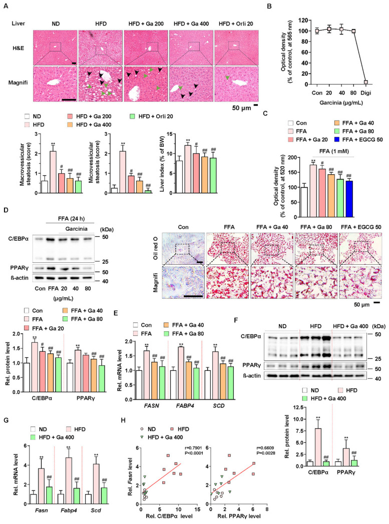
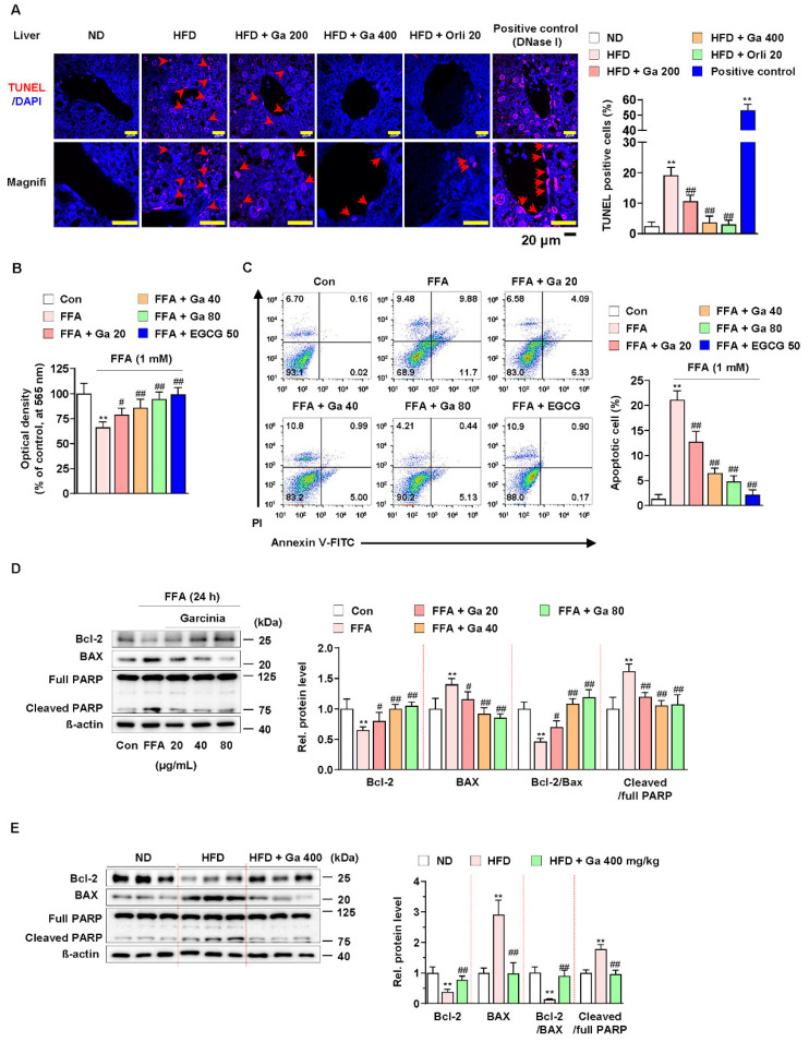
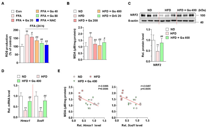
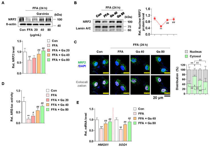
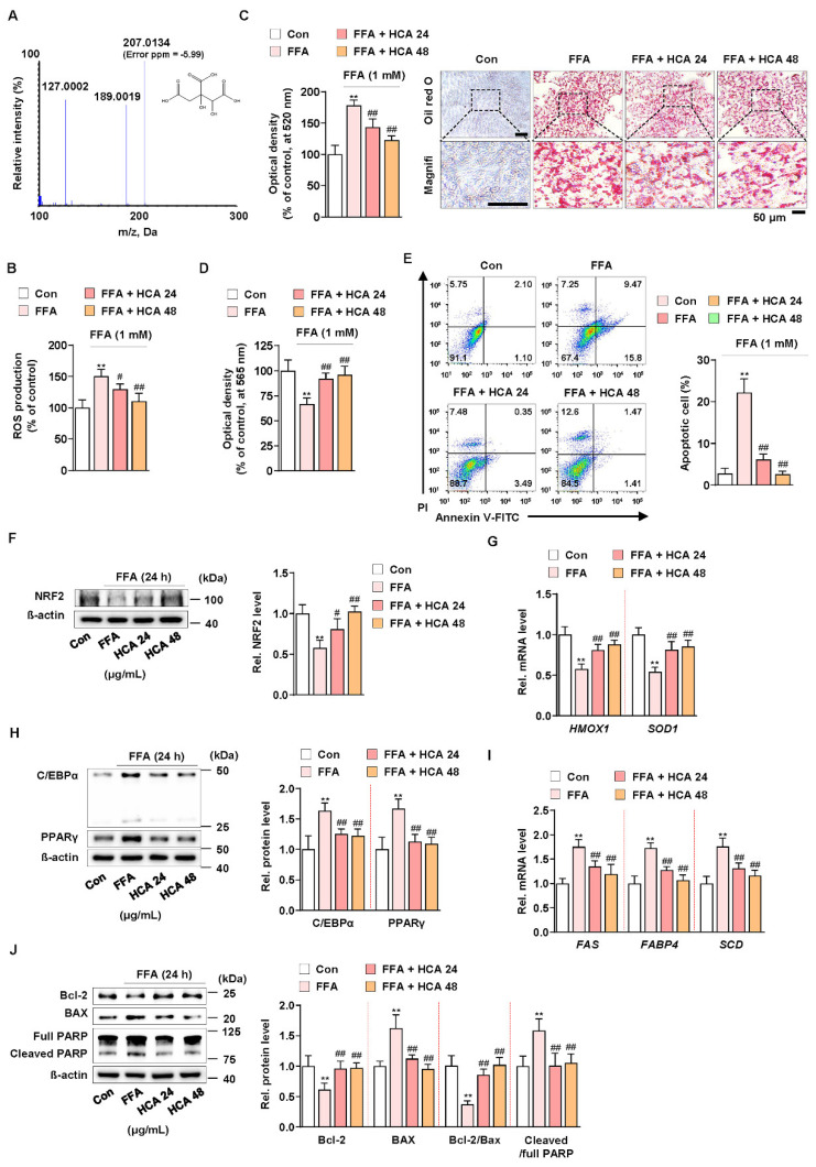
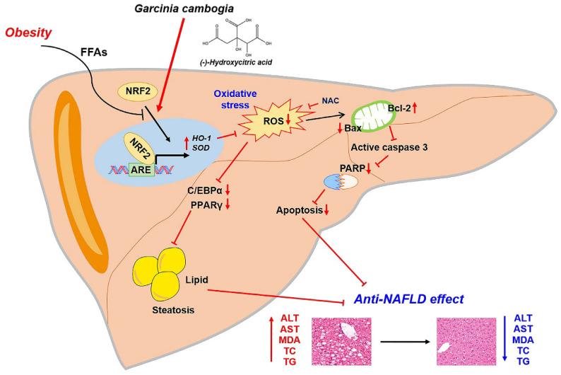
Excessive free fatty acids (FFAs) causes reactive oxygen species (ROS) generation and non-alcoholic fatty liver disease (NAFLD) development. Garcinia cambogia (G. cambogia) is used as an anti-obesity supplement, and its protective potential against NAFLD has been investigated. This study aims to present the therapeutic effects of G. cambogia on NAFLD and reveal underlying mechanisms. High-fat diet (HFD)-fed mice were administered G. cambogia for eight weeks, and steatosis, apoptosis, and biochemical parameters were examined in vivo. FFA-induced HepG2 cells were treated with G. cambogia, and lipid accumulation, apoptosis, ROS level, and signal alterations were examined. The results showed that G. cambogia inhibited HFD-induced steatosis and apoptosis and abrogated abnormalities in serum chemistry. G. cambogia increased in NRF2 nuclear expression and activated antioxidant responsive element (ARE), causing induction of antioxidant gene expression. NRF2 activation inhibited FFA-induced ROS production, which suppressed lipogenic transcription factors, C/EBPα and PPARγ. Moreover, the ability of G. cambogia to inhibit ROS production suppressed apoptosis by normalizing the Bcl-2/BAX ratio and PARP cleavage. Lastly, these therapeutic effects of G. cambogia were due to hydroxycitric acid (HCA). These findings provide new insight into the mechanism by which G. cambogia regulates NAFLD progression.

Keywords: Garcinia cambogia; NRF2; antioxidant; apoptosis; hydroxycitric acid; non-alcoholic fatty liver disease (NAFLD); reactive oxygen species; steatosis.


Conflict of interest statement

The authors declare no conflict of interest.

Figures

Figure 1
Figure 1
Anti-NALFD effect of Garcinia cambogia in HFD-fed mice reducing hepatic steatosis. (A) Representative images showing the effect of G. cambogia (Ga, 200 and 400 mg/kg) on HFD-induced hepatic steatosis. H&E staining of liver tissues was performed. The scores of macrovesicular (black arrow) and microvesicular (green arrow) steatosis and liver index were quantified, as described in the Methods section. Normal diet (ND)-fed mice were used as the negative control for high levels of fat accumulation, and orlistat (20 mg/kg) was used as the positive control for anti-obesity and anti-steatosis effects (n = 7 per group). Scale bars: 50 μm. Magnifi: magnification. ** p < 0.01 vs. ND-fed mice, # p < 0.05 and ## p < 0.01 vs. HFD-fed mice. (B) MTT assay showing the effect of G. cambogia (20–80 μg/mL) on cell viability. HepG2 cells were treated with the indicated concentration of G. cambogia and digitonin (100 μg/mL, positive control) for 24 h (n = 5 per group). (C) oil red O assay and representative images showing the effect of G. cambogia (20–80 μg/mL) on free fatty acid (1 mM FFA)-induced lipid accumulation in HepG2 cells. FFA-induced HepG2 cells were treated with G. cambogia and EGCG (50 μM, positive control) for 24 h (n = 5 per group). Scale bar: 50 μm. Magnifi: magnification. ** p < 0.01 vs. Con, # p < 0.05 and ## p < 0.01 vs. FFA. Effect of G. cambogia (20–80 μg/mL) on (D) C/EBPα and PPARγ expression and (E) transcript levels of FASN, FABP4, and SCD in FFA-treated HepG2 cells (d, n = 4 per group; e, n = 5 per group). Effect of G. cambogia on (F) C/EBPα and PPARγ expression and (G) Fasn, Fabp4, and Scd transcript levels of liver tissues from ND-fed, HFD-fed, and HFD-fed mice administered a high dose of G. cambogia (400 mg/kg) (n = 6 per group). (H) Correlations between Fasn transcript and C/EBPα and PPARγ protein levels in liver tissues (n = 6 per group). Each point represents one sample. Data are mean ± S.D.
Figure 2
Figure 2
Effect of Garcinia cambogia on HFD-induced apoptosis in the liver. (A) Representative images of TUNEL staining and quantification data for the number of TUNEL-positive cells in HFD-induced liver tissues. Positive control: DNase I (30 U) treated group. Scale bar: 20 μm. (n = 6 per group). Magnifi: magnification. ** p < 0.01 vs. ND-fed mice, ## p < 0.01 vs. HFD-fed mice. (B) MTT assay showing the effect of G. cambogia on FFA-induced cell viability. Cells were treated with G. cambogia (20–80 μg/mL) and EGCG (50 μM) for 24 h (n = 5 per group). ** p < 0.01 vs. Con, # p < 0.05 and ## p < 0.01 vs. FFA. (C) Effect of G. cambogia on apoptosis in FFA-treated HepG2 cells. Cells were treated with G. cambogia (20–80 μg/mL) and EGCG (50 μM) for 24 h (n = 4 per group). The numbers of negatively and positively stained cells are expressed as percentages of the total number of cells. (D) Effect of G. cambogia on Bcl-2 and BAX expression and PARP cleavage in FFA-treated HepG2 cells (n = 4 per group). (E) Effect of G. cambogia on Bcl-2 and BAX expression and PARP cleavage of liver tissues from ND-fed, HFD-fed, and HFD-fed mice administered a high dose of G. cambogia (400 mg/kg) (n = 6 per group). Data are mean ± S.D.
Figure 3
Figure 3
Effect of Garcinia cambogia on HFD- and FFA-induced ROS production, and NRF2 and downstream gene activation. (A) Effect of G. cambogia on ROS level in FFA-treated HepG2 cells. Cells were treated with G. cambogia (20–80 μg/mL) or NAC (5 mM, positive control of antioxidant) for 24 h. ROS production was measured using H2DCFDA (n = 5 per group). ** p < 0.01 vs. Con, # p < 0.05 and ## p < 0.01 vs. FFA. (B) Effect of G. cambogia on malondialdehyde (MDA, indicating the intensity of lipid peroxidation) level of serum in HFD-fed mice (n = 7 per group). Effect of G. cambogia on (C) NRF2 expression and (D) Hmox1 and Sod1 transcript levels of liver tissues from ND-fed, HFD-fed, and HFD-fed mice administered a high dose of G. cambogia (400 mg/kg) (n = 6 per group). (E) Correlations between MDA level and Hmox1 and Sod1 transcript levels in liver tissues (n = 6 per group). Each point represents one sample. ** p < 0.01 vs. ND-fed mice, # p < 0.05 and ## p < 0.01 vs. HFD-fed mice. Data are mean ± S.D.
Figure 4
Figure 4
Effect of Garcinia cambogia on NRF2-ARE activation in FFA-induced HepG2 cells. (A) Effect of G. cambogia (20–80 μg/mL) on NRF2 expression in FFA-treated HepG2 cells (n = 4 per group). (B) Effect of G. cambogia on NRF2 nuclear expression in FFA-treated HepG2 cells. Cells were treated with G. cambogia (40 and 80 μg/mL) for 12 h, and cells were fractionated into nucleus compartment as described in the Methods section (n = 4 per group). (C) Representative immunofluorescence images of NRF2 in FFA-treated HepG2 cells. Colocalized FITC (i.e., NRF2) and DAPI (i.e., nuclei) were quantified using ImageJ software, and relative intensities in the nuclear and cytoplasmic fractions were expressed as a histogram. Scale bars: 20 μm (n = 5 per group). (D) Effect of G. cambogia on ARE promoter activity in FFA-treated HepG2 cells. ARE-luc vector-transfected HepG2 cells were cotreated FFA and G. cambogia (20–80 μg/mL) for 12 h and detected ARE promoter activity (n = 6 per group). (E) Effect of G. cambogia (20–80 μg/mL) on the transcript levels of HMOX1 and SOD1 in FFA-treated HepG2 cells (n = 5 per group). ** p < 0.01 vs. Con, # p < 0.05 and ## p < 0.01 vs. FFA. Data are mean ± S.D.
Figure 5
Figure 5
Effect of hydroxycitric acid on FFA-induced ROS production, lipid accumulation, and apoptosis in HepG2 cells. (A) MS/MS spectrum of hydroxycitric acid [M − H]. The parent ion was the deprotonated [M − H] ion at m/z 207.0 and the most abundant ion in product ion scan mode. (B) ROS measurement using H2DCFDA in FFA-treated HepG2 cells treated with HCA (24 and 48 μg/mL) (n = 5 per group). (C) Oil red O assay and representative images showing the effect of HCA (24 and 48 μg/mL) on FFA-induced lipid accumulation in HepG2 cells. Cells were treated with HCA (24 and 48 μg/mL) for 24 h (n = 5 per group). Scale bar: 50 μm. Magnifi: magnification. (D) MTT assay showing the effect of HCA (24 and 48 μg/mL) on FFA-induced cell viability. Cells were treated with HCA (24 and 48 μg/mL) for 24 h (n = 5 per group). (E) Effect of HCA (24 and 48 μg/mL) on apoptosis in FFA-treated HepG2 cells. Cells were treated with HCA (24 and 48 μg/mL) for 24 h (n = 4 per group). The numbers of negatively and positively stained cells are expressed as percentages of the total number of cells. Effect of HCA (24 and 48 μg/mL) on the levels of (F) NRF2 expression, (G) HMOX1 and SOD1 transcriptions, (H) C/EBPα and PPARγ expression, (I) FAS, FABP4 and SCD transcriptions, and (J) Bcl-2 and BAX expression and PARP cleavage in FFA-treated HepG2 cells (n = 4 per group in f, h, and j; n = 5 per group in G,I). ** p < 0.01 vs. Con, # p < 0.05 and ## p < 0.01 vs. FFA. Data are mean ± S.D.
Figure 6
Figure 6
Proposed mechanism of the protective effect of G. cambogia against NAFLD. G. cambogia inhibited HFD-induced NAFLD by activating the NRF2-ARE-mediated antioxidant defense system, leading to inhibition of hepatic steatosis and apoptosis in the liver.

References

    1. Hill J.O., Wyatt H.R., Peters J.C. Energy balance and obesity. Circulation. 2012;126:126–132. doi: 10.1161/CIRCULATIONAHA.111.087213. - DOI - PMC - PubMed
    1. Paschos P., Paletas K. Non alcoholic fatty liver disease and metabolic syndrome. Hippokratia. 2009;13:9–19. - PMC - PubMed
    1. Younossi Z.M. Non-alcoholic fatty liver disease—A global public health perspective. J. Hepatol. 2019;70:531–544. doi: 10.1016/j.jhep.2018.10.033. - DOI - PubMed
    1. Fabbrini E., Sullivan S., Klein S. Obesity and nonalcoholic fatty liver disease: Biochemical, metabolic, and clinical implications. Hepatology. 2010;51:679–689. doi: 10.1002/hep.23280. - DOI - PMC - PubMed
    1. Parthasarathy G., Revelo X., Malhi H. Pathogenesis of Nonalcoholic Steatohepatitis: An Overview. Hepatol. Commun. 2020;4:478–492. doi: 10.1002/hep4.1479. - DOI - PMC - PubMed