. 2021 Aug 11;10(8):1274.
doi: 10.3390/antiox10081274.

The Interplay of Mitochondrial Oxidative Stress and Endoplasmic Reticulum Stress in Cardiovascular Fibrosis in Obese Rats

Affiliations

The Interplay of Mitochondrial Oxidative Stress and Endoplasmic Reticulum Stress in Cardiovascular Fibrosis in Obese Rats

Francisco V Souza-Neto et al. Antioxidants (Basel). .

Abstract

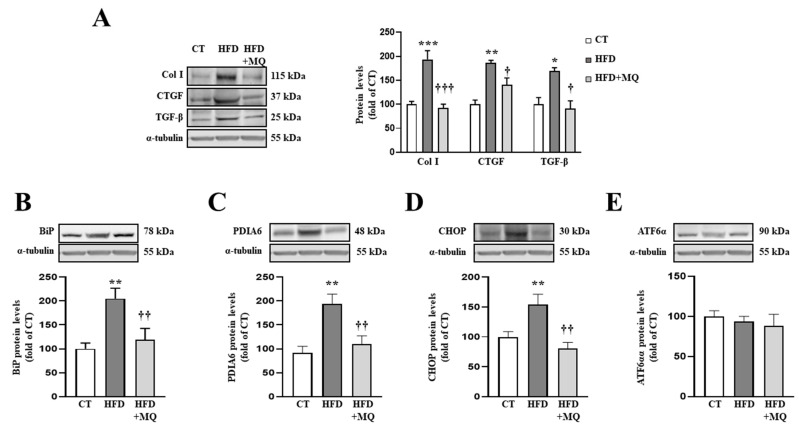
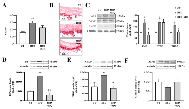
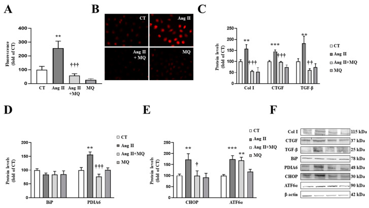
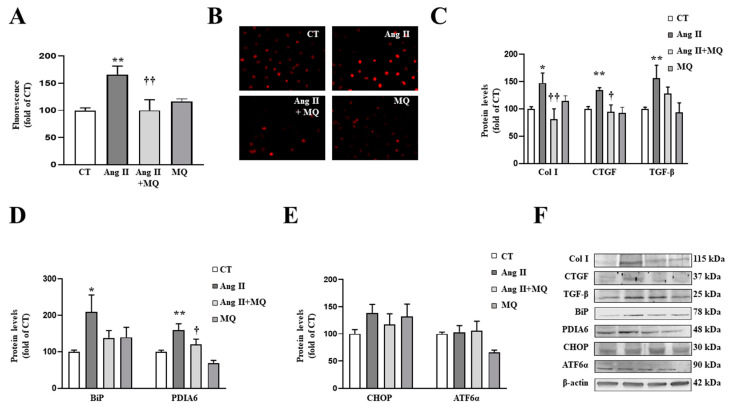
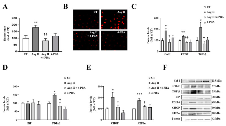
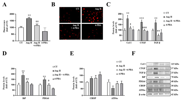
We have evaluated the role of mitochondrial oxidative stress and its association with endoplasmic reticulum (ER) stress activation in the progression of obesity-related cardiovascular fibrosis. MitoQ (200 µM) was orally administered for 7 weeks to male Wistar rats that were fed a high-fat diet (HFD, 35% fat) or a control diet (CT, 3.5% fat). Obese animals presented cardiovascular fibrosis accompanied by increased levels of extracellular matrix proteins and profibrotic mediators. These alterations were associated with ER stress activation characterized by enhanced levels (in heart and aorta vs. CT group, respectively) of immunoglobulin binding protein (BiP; 2.1-and 2.6-fold, respectively), protein disulfide-isomerase A6 (PDIA6; 1.9-fold) and CCAAT-enhancer-binding homologous protein (CHOP; 1.5- and 1.8-fold, respectively). MitoQ treatment was able to prevent (p < 0.05) these modifications at cardiac and aortic levels. MitoQ (5 nM) and the ER stress inhibitor, 4-phenyl butyric acid (4 µM), were able to block the prooxidant and profibrotic effects of angiotensin II (Ang II, 10-6 M) in cardiac and vascular cells. Therefore, the data show a crosstalk between mitochondrial oxidative stress and ER stress activation, which mediates the development of cardiovascular fibrosis in the context of obesity and in which Ang II can play a relevant role.

Keywords: cardiovascular fibrosis; endoplasmic reticulum stress; mitochondrial oxidative stress; obesity.


Conflict of interest statement

The authors declare no conflict of interest.

Figures

Figure 1
Figure 1
Mitochondrial oxidative stress mediates the increase in extracellular matrix proteins and endoplasmic reticulum stress at cardiac level. Protein levels of (A) collagen type I (Col I), connective tissue growth factor (CTGF) and transforming growth factor-beta (TGF-β); (B) immunoglobulin binding protein (BiP); (C) protein disulfide isomerase family A member 6 (PDIA6); (D) CCAAT-enhancer-binding protein homologous protein (CHOP); (E) activating transcription factor 6-alpha (ATF6α) in heart tissue from control rats fed a normal chow (CT) and rats fed a high-fat diet (HFD) treated with vehicle or with the mitochondrial antioxidant MitoQ (MQ; 200 µM). Bars graphs represent the mean ± SEM of 6–8 animals normalized for α-tubulin. * p < 0.05, ** p < 0.01, *** p < 0.001 vs. control group. † p < 0.05, †† p < 0.01, ††† p < 0.001 vs. HFD group.
Figure 2
Figure 2
Mitochondrial oxidative stress mediates vascular fibrosis and endoplasmic reticulum stress at aortic level. (A) Quantification of collagen volume fraction and (B) representative microphotographs of aortic sections staining with picrosirius red. Protein levels of (C) collagen type I (Col I), connective tissue growth factor (CTGF) and transforming growth factor-beta (TGF-β); (D) immunoglobulin binding protein (BiP); (E) CCAAT-enhancer-binding protein homologous protein (CHOP); (F) activating transcription factor 6-alpha (ATF6α) in aortic tissue from control rats fed a normal chow (CT) and rats fed a high-fat diet (HFD) treated with vehicle or with the mitochondrial antioxidant MitoQ (MQ; 200 µM). Scale bar: 50 µm. Bars graphs represent the mean ± SEM of 6–8 animals normalized for α-tubulin. * p < 0.05, ** p < 0.01 vs. control group. † p < 0.05, †† p < 0.01 vs. HFD group.
Figure 3
Figure 3
Mitochondrial oxidative stress mediates the prooxidant, profibrotic and the endoplasmic reticulum stress activation induced by angiotensin II (Ang II) in cardiac fibroblasts. (A,B) Effect of the mitochondrial antioxidant MitoQ (MQ; 5 nM) on superoxide anion production. Quantification of cells labelled with the oxidative dye dihydroethidium (A) and representative microphotographs (magnification 40×) (B). Protein levels of (C) collagen type I (Col I), connective tissue growth factor (CTGF) and transforming growth factor-beta (TGF-β); (D) immunoglobulin binding protein (BiP) and protein disulfide isomerase family A member 6 (PDIA6); (E) CCAAT-enhancer-binding protein homologous protein (CHOP) and activating transcription factor 6-alpha (ATF6α) in cardiac fibroblasts treated with Ang II (10−6 M) for 24 h in the presence or in the absence of MQ. (F) Representative blots for protein expressions. Bars graphs represent the mean ± SEM of MS four to six assays normalized for β-actin. ** p < 0.01, *** p < 0.001 vs. control cells. † p < 0.05, †† p < 0.01, ††† p < 0.001 vs. Ang II-treated cardiac fibroblasts.
Figure 4
Figure 4
Mitochondrial oxidative stress mediates the prooxidant, profibrotic and the endoplasmic reticulum stress activation induced by angiotensin II (Ang II) in vascular smooth muscle cells (VSMCs). (A,B) Effects of the mitochondrial antioxidant MitoQ (MQ; 5 nM) on superoxide anion production. Quantification of cells labelled with the oxidative dye dihydroethidium (A) and representative microphotographs (magnification 40×) (B). Protein levels of (C) collagen type I (Col I), connective tissue growth factor (CTGF) and transforming growth factor-beta (TGF-β); (D) immunoglobulin binding protein (BiP) and protein disulfide isomerase family A member 6 (PDIA6); (E) CCAAT-enhancer-binding protein homologous protein (CHOP) and activating transcription factor 6-alpha (ATF6α) in VSMCs treated with Ang II (10−6 M) for 24 h in the presence or in the absence of MQ. (F) Representative blots for protein expressions. Bars graphs represent the mean ± SEM of four to six assays normalized for β-actin. * p < 0.05, ** p < 0.01, vs. control cells. † p < 0.05, †† p < 0.01, vs. Ang II-treated VSMCs.
Figure 5
Figure 5
Endoplasmic reticulum stress mediates the prooxidant and profibrotic effects induced by angiotensin II (Ang II) in cardiac fibroblasts. (A,B) Effect of 4-phenylbutyrate acid (4-PBA; 4 µM) on superoxide anion production. Quantification of cells labelled with the oxidative dye dihydroethidium (A) and representative microphotographs (magnification 40×) (B). Protein levels of (C) collagen type I (Col I), connective tissue growth factor (CTGF) and transforming growth factor-beta (TGF-β); (D) immunoglobulin binding protein (BiP) and protein disulfide isomerase family A member 6 (PDIA6); (E) CCAAT-enhancer-binding protein homologous protein (CHOP) and activating transcription factor 6-alpha (ATF6α) in cardiac fibroblasts treated with Ang II (10−6 M) for 24 h in the presence or in the absence of 4-PBA. (F) Representative blots for protein expressions. Bars graphs represent the mean ± SEM of four to six assays normalized for β-actin. * p < 0.05, ** p < 0.01, *** p < 0.001 vs. control cells. † p < 0.05, †† p < 0.01 vs. Ang II-treated cardiac fibroblasts.
Figure 6
Figure 6
Endoplasmic reticulum stress mediates the prooxidant and profibrotic effects induced by Angiotensin II (Ang II) in vascular smooth muscle cells (VSMCs). (A,B) Effect of 4-phenylbutyrate acid (4-PBA; 4 µM) on superoxide anion production. Quantification of cells labelled with the oxidative dye dihydroethidium (A) and representative microphotographs (magnification 40×) (B). Protein levels of (C) collagen type I (Col I), connective tissue growth factor (CTGF) and transforming growth factor-beta (TGF-β); (D) immunoglobulin binding protein (BiP) and protein disulfide isomerase family A member 6 (PDIA6); (E) CCAAT-enhancer-binding protein homologous protein (CHOP) and activating transcription factor 6-alpha (ATF6α) in VSMCs treated with Ang II (10−6 M) for 24 h. (F) Representative blots for protein expressions. Bars graphs represent the mean ± SEM of four to six assays normalized for β-actin. ** p < 0.01, *** p < 0.001 vs. control cells. † p < 0.05, †† p < 0.01, ††† p < 0.001 vs. Ang II-treated VSMCs.

References

    1. Kong P., Christia P., Frangogiannis N.G. The pathogenesis of cardiac fibrosis. Cell Mol. Life Sci. 2014;71:549–574. doi: 10.1007/s00018-013-1349-6. - DOI - PMC - PubMed
    1. Czubryt M.P. Cardiac Fibroblast to Myofibroblast Phenotype Conversion-An Unexploited Therapeutic Target. J. Cardiovasc. Dev. Dis. 2019;6:28. doi: 10.3390/jcdd6030028. - DOI - PMC - PubMed
    1. Kirkman D.L., Robinson A.T., Rossman M.J., Seals D.R., Edwards D.G. Mitochondrial contributions to vascular endothelial dysfunction, arterial stiffness, and cardiovascular diseases. Am. J. Physiol. Heart Circ. Physiol. 2021;320:H2080–H2100. doi: 10.1152/ajpheart.00917.2020. - DOI - PMC - PubMed
    1. Cavalera M., Wang J., Frangogiannis N.G. Obesity, metabolic dysfunction, and cardiac fibrosis: Pathophysiological pathways, molecular mechanisms, and therapeutic opportunities. Transl. Res. 2014;164:323–335. doi: 10.1016/j.trsl.2014.05.001. - DOI - PMC - PubMed
    1. Martinez-Martinez E., Jurado-Lopez R., Valero-Munoz M., Bartolome M.V., Ballesteros S., Luaces M., Briones A.M., Lopez-Andres N., Miana M., Cachofeiro V. Leptin induces cardiac fibrosis through galectin-3, mTOR and oxidative stress: Potential role in obesity. J. Hypertens. 2014;32:1104–1114; discussion 1114. doi: 10.1097/HJH.0000000000000149. - DOI - PubMed