. 2021 Jul 21;11(8):960.
doi: 10.3390/brainsci11080960.

The Right Hemisphere Is Responsible for the Greatest Differences in Human Brain Response to High-Arousing Emotional versus Neutral Stimuli: A MEG Study

Affiliations

The Right Hemisphere Is Responsible for the Greatest Differences in Human Brain Response to High-Arousing Emotional versus Neutral Stimuli: A MEG Study

Mina Kheirkhah et al. Brain Sci. .

Abstract

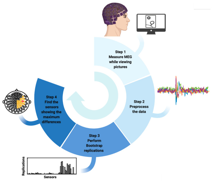
Studies investigating human brain response to emotional stimuli-particularly high-arousing versus neutral stimuli-have obtained inconsistent results. The present study was the first to combine magnetoencephalography (MEG) with the bootstrapping method to examine the whole brain and identify the cortical regions involved in this differential response. Seventeen healthy participants (11 females, aged 19 to 33 years; mean age, 26.9 years) were presented with high-arousing emotional (pleasant and unpleasant) and neutral pictures, and their brain responses were measured using MEG. When random resampling bootstrapping was performed for each participant, the greatest differences between high-arousing emotional and neutral stimuli during M300 (270-320 ms) were found to occur in the right temporo-parietal region. This finding was observed in response to both pleasant and unpleasant stimuli. The results, which may be more robust than previous studies because of bootstrapping and examination of the whole brain, reinforce the essential role of the right hemisphere in emotion processing.

Keywords: bootstrapping; emotion; magnetoencephalography (MEG); right hemisphere.


Conflict of interest statement

C.A.Z. is listed as a co-inventor on a patent for the use of ketamine in major depression and suicidal ideation; as a co-inventor on a patent for the use of (2R,6R)-hydroxynorketamine, (S)-dehydronorketamine, and other stereoisomeric dehydroxylated and hydroxylated metabolites of (R,S)-ketamine metabolites in the treatment of depression and neuropathic pain; and as a co-inventor on a patent application for the use of (2R,6R)-hydroxynorketamine and (2S,6S)-hydroxynorketamine in the treatment of depression, anxiety, anhedonia, suicidal ideation, and post-traumatic stress disorders. He has assigned his patent rights to the U.S. government but will share a percentage of any royalties that may be received by the government. All other authors have no conflict of interest to disclose, financial or otherwise.

Figures

Figure 1
Figure 1
Workflow of the proposed algorithm to find the MEG sensors showing greater differences in human brain response to high-arousing emotional stimuli compared to neutral stimuli.
Figure 2
Figure 2
Selection of sensors in bootstrapping replications. The bar plots represent the sensors that expressed the highest differences in each participant’s brain response to highly arousing pleasant (left) and unpleasant (right) emotional pictures compared to neutral pictures during the M300 time interval detected in each bootstrap replication. The Y-axis indicates the 25,000 bootstrap replications. The X-axis shows the 102 magnetometers encompassing the whole brain.
Figure 2
Figure 2
Selection of sensors in bootstrapping replications. The bar plots represent the sensors that expressed the highest differences in each participant’s brain response to highly arousing pleasant (left) and unpleasant (right) emotional pictures compared to neutral pictures during the M300 time interval detected in each bootstrap replication. The Y-axis indicates the 25,000 bootstrap replications. The X-axis shows the 102 magnetometers encompassing the whole brain.
Figure 3
Figure 3
Selected sensors showing the highest differences in brain responses to pleasant and unpleasant versus neutral pictures for each participant. The plots depict the magnetoencephalography (MEG) sensors across the entire head (small black circles). Highlighted sensors (in orange and red) indicate the sensors selected by bootstrapping, showing the largest differences in brain response to pleasant and unpleasant versus neutral pictures for each participant. The highlighted orange sensors are those that were selected by more than 20% of bootstrapping replications, and the red sensors are those that showed significantly higher responses to high-arousing emotional pictures versus neutral pictures within 20% of bootstrapping replications.
Figure 4
Figure 4
Forest plot for all selected sensors by bootstrapping over all participants. Vertical lines show the 95% confidence intervals for the selected sensors based on 25,000 bootstrapping replications for all participants, with the median values represented by black dots. Orange lines show the most frequently selected sensors for all participants with the highest median and the highest lower limits in confidence intervals. The bottom graphs depict the location of the selected sensors (orange lines in the top figures) which are located in the right temporal and parietal regions.

References

    1. Ahern G.L., Schwartz G.E. Differential lateralization for positive versus negative emotion. Neuropsychologia. 1979;17:693–698. doi: 10.1016/0028-3932(79)90045-9. - DOI - PubMed
    1. Borod J.C., Cicero B.A., Obler L.K., Welkowitz J., Erhan H.M., Santschi C., Grunwald I.S., Agosti R.M., Whalen J.R. Right hemisphere emotional perception: Evidence across multiple channels. Neuropsychology. 1998;12:446–458. doi: 10.1037/0894-4105.12.3.446. - DOI - PubMed
    1. Gainotti G. Emotional behavior and hemispheric side of the lesion. Cortex. 1972;8:41–55. doi: 10.1016/S0010-9452(72)80026-1. - DOI - PubMed
    1. Gainotti G. Face familiarity feelings, the right temporal lobe and the possible underlying neural mechanisms. Brain Res. Rev. 2007;56:214–235. doi: 10.1016/j.brainresrev.2007.07.009. - DOI - PubMed
    1. Gainotti G. Unconscious processing of emotions and the right hemisphere. Neuropsychologia. 2012;50:205–218. doi: 10.1016/j.neuropsychologia.2011.12.005. - DOI - PubMed

LinkOut - more resources