Review
. 2021 Jul 31;11(8):1129.
doi: 10.3390/biom11081129.

Cranial Suture Mesenchymal Stem Cells: Insights and Advances

Affiliations
Review

Cranial Suture Mesenchymal Stem Cells: Insights and Advances

Bo Li et al. Biomolecules. .

Abstract

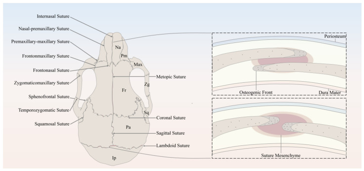
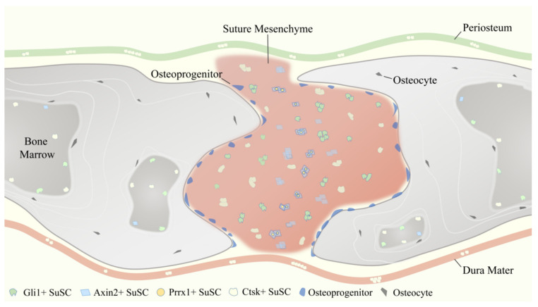
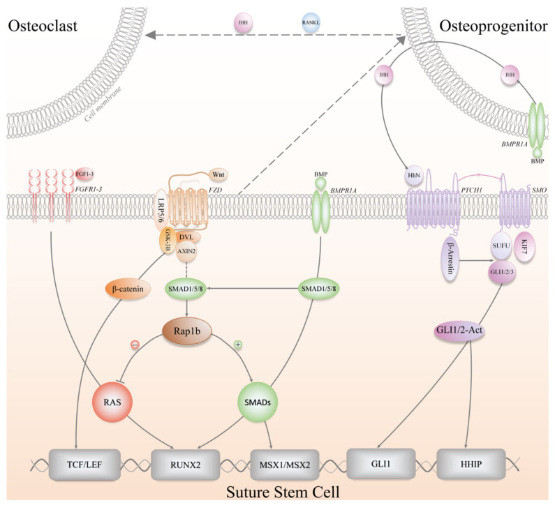
The cranial bones constitute the protective structures of the skull, which surround and protect the brain. Due to the limited repair capacity, the reconstruction and regeneration of skull defects are considered as an unmet clinical need and challenge. Previously, it has been proposed that the periosteum and dura mater provide reparative progenitors for cranial bones homeostasis and injury repair. In addition, it has also been speculated that the cranial mesenchymal stem cells reside in the perivascular niche of the diploe, namely, the soft spongy cancellous bone between the interior and exterior layers of cortical bone of the skull, which resembles the skeletal stem cells' distribution pattern of the long bone within the bone marrow. Not until recent years have several studies unraveled and validated that the major mesenchymal stem cell population of the cranial region is primarily located within the suture mesenchyme of the skull, and hence, they are termed suture mesenchymal stem cells (SuSCs). Here, we summarized the characteristics of SuSCs, this newly discovered stem cell population of cranial bones, including the temporospatial distribution pattern, self-renewal, and multipotent properties, contribution to injury repair, as well as the signaling pathways and molecular mechanisms associated with the regulation of SuSCs.

Keywords: Axin2; Ctsk; Gli1; Prrx1; cranial sutures; injury repair; mesenchymal stem cells.


Conflict of interest statement

The authors declare no conflict of interest.

Figures

Figure 1
Figure 1
Schematic of the murine skull in the dorsal view, depicting the anatomy of cranial bones and sutures. Na, Nasal; Pm, Premaxilla; Max, Maxilla; Zg, Zygomatic; Sq, Squamosal; Fr, Frontal; Pa, Parietal; Ip, Interparietal.
Figure 2
Figure 2
Schematic of different types of craniosynostosis, including trigonocephaly, scaphocephaly, anterior plagiocephaly, posterior plagiocephaly, and brachycephaly.
Figure 3
Figure 3
Temporospatial distribution pattern of four representative suture mesenchymal stem cells (SuSCs) subpopulations, including Gli1+ SuSCs, Axin2+ SuSCs, Prrx1+ SuSCs, and Ctsk+ SuSCs.
Figure 4
Figure 4
The signaling pathways involved in the regulation of SuSCs, including Wnt (Wnt/β-catenin), FGF (Fibroblast growth factor), BMP (Bone morphogenetic protein), Hh (Hedgehog) signaling pathways. IHH, Indian hedgehog; RANKL, Receptor activator of nuclear factor-κB ligand; BMPR1A, Bone morphogenetic protein receptor type 1A; FGFR, Fibroblast growth factor receptor; FZD, Frizzled; LRP5/6, Low-density lipoprotein-related receptors 5/6; DVL, Dishevelled; GSK-3B, Glycogen synthase kinase 3-beta; PTCH1, Patched 1; SMO, Smoothened; SUFU, Suppressor of fused; KIF7, Kinesin family member 7; TCF/LEF, T-cell factor/Lymphoid-enhancer factor; RUNX2, Runt-related transcription factor 2; MSX1/2, Msh homeobox 1/2; HHIP, Hedgehog interacting protein.

References

    1. Zhao H., Feng J., Ho T.V., Grimes W., Urata M., Chai Y. The suture provides a niche for mesenchymal stem cells of craniofacial bones. Nat. Cell Biol. 2015;17:386–396. doi: 10.1038/ncb3139. - DOI - PMC - PubMed
    1. Maruyama T., Jeong J., Sheu T.J., Hsu W. Stem cells of the suture mesenchyme in craniofacial bone development, repair and regeneration. Nat. Commun. 2016;7:10526. doi: 10.1038/ncomms10526. - DOI - PMC - PubMed
    1. Wilk K., Yeh S.C.A., Mortensen L.J., Ghaffarigarakani S., Lombardo C.M., Bassir S.H., Aldawood Z.A., Lin C.P., Intini G. Postnatal Calvarial Skeletal Stem Cells Expressing PRX1 Reside Exclusively in the Calvarial Sutures and Are Required for Bone Regeneration. Stem Cell Rep. 2017;8:933–946. doi: 10.1016/j.stemcr.2017.03.002. - DOI - PMC - PubMed
    1. Debnath S., Yallowitz A.R., McCormick J., Lalani S., Zhang T., Xu R., Li N., Liu Y., Yang Y.S., Eiseman M., et al. Discovery of a periosteal stem cell mediating intramembranous bone formation. Nature. 2018;562:133–139. doi: 10.1038/s41586-018-0554-8. - DOI - PMC - PubMed
    1. Chan C.K.F., Seo E.Y., Chen J.Y., Lo D., McArdle A., Sinha R., Tevlin R., Seita J., Vincent-Tompkins J., Wearda T., et al. Identification and specification of the mouse skeletal stem cell. Cell. 2015;160:285–298. doi: 10.1016/j.cell.2014.12.002. - DOI - PMC - PubMed

Publication types

MeSH terms