. 2021 Jul 21;9(8):851.
doi: 10.3390/biomedicines9080851.

Noble Metals for Modern Implant Materials: MOCVD of Film Structures and Cytotoxical, Antibacterial, and Histological Studies

Affiliations

Noble Metals for Modern Implant Materials: MOCVD of Film Structures and Cytotoxical, Antibacterial, and Histological Studies

Svetlana I Dorovskikh et al. Biomedicines. .

Abstract

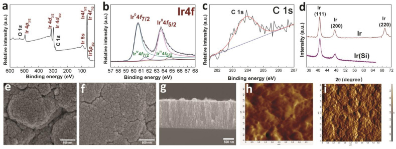
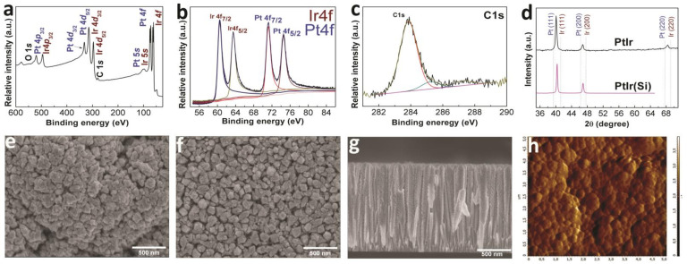
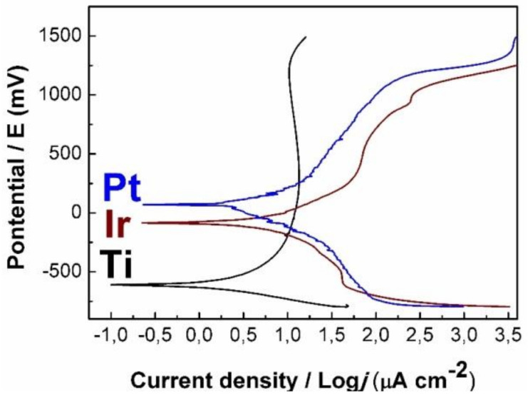
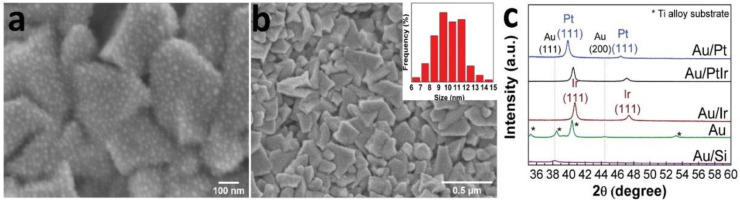
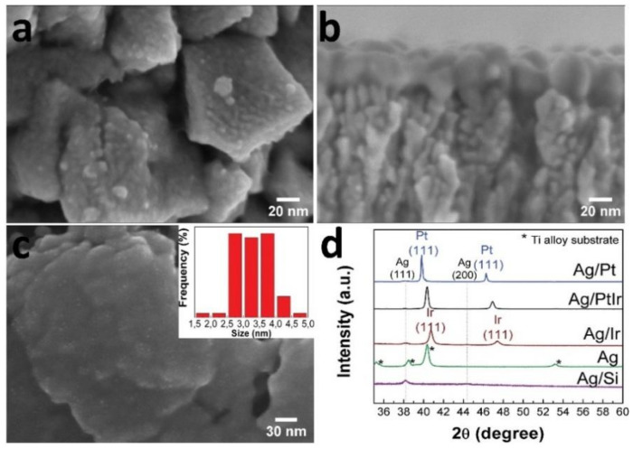
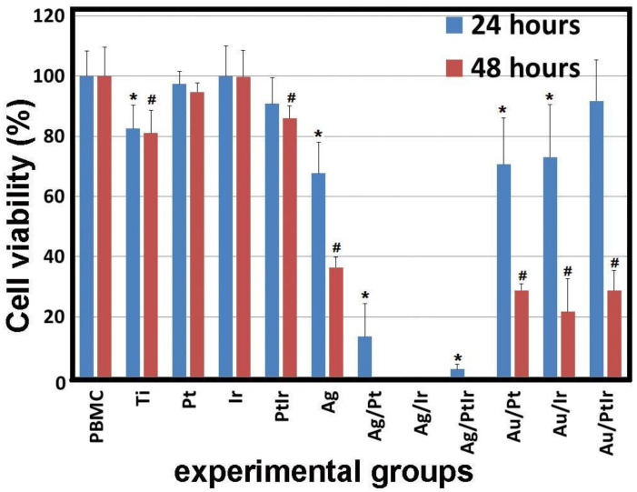
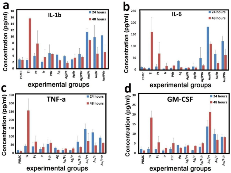
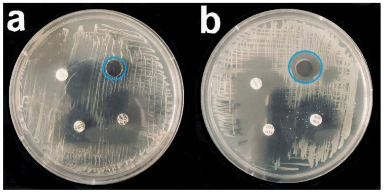
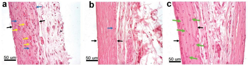
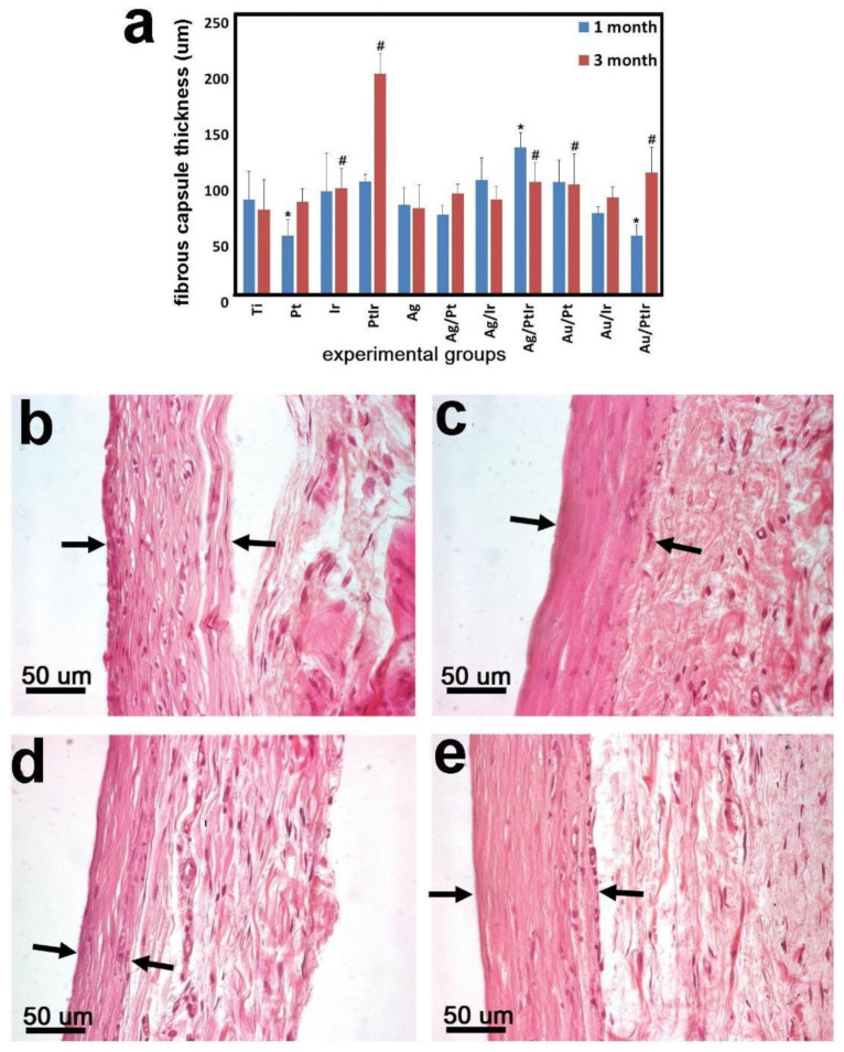
This work is aimed at developing the modification of the surface of medical implants with film materials based on noble metals in order to improve their biological characteristics. Gas-phase transportation methods were proposed to obtain such materials. To determine the effect of the material of the bottom layer of heterometallic structures, Ir, Pt, and PtIr coatings with a thickness of 1.4-1.5 μm were deposited by metal-organic chemical vapor deposition (MOCVD) on Ti6Al4V alloy discs. Two types of antibacterial components, namely, gold nanoparticles (AuNPs) and discontinuous Ag coatings, were deposited on the surface of these coatings. AuNPs (11-14 nm) were deposited by a pulsed MOCVD method, while Ag films (35-40 nm in thickness) were obtained by physical vapor deposition (PVD). The cytotoxic (24 h and 48 h, toward peripheral blood mononuclear cells (PBMCs)) and antibacterial (24 h) properties of monophase (Ag, Ir, Pt, and PtIr) and heterophase (Ag/Pt, Ag/Ir, Ag/PtIr, Au/Pt, Au/Ir, and Au/PtIr) film materials deposited on Ti-alloy samples were studied in vitro and compared with those of uncoated Ti-alloy samples. Studies of the cytokine production by PBMCs in response to incubation of the samples for 24 and 48 h and histological studies at 1 and 3 months after subcutaneous implantation in rats were also performed. Despite the comparable thickness of the fibrous capsule after 3 months, a faster completion of the active phase of encapsulation was observed for the coated implants compared to the Ti alloy analogs. For the Ag-containing samples, growth inhibition of S. epidermidis, S. aureus, Str. pyogenes, P. aeruginosa, and Ent. faecium was observed.

Keywords: biochemical and cytokine blood composition; chemical vapor deposition; cytological; gold; histological study; iridium; platinum; silver; thin films and nanoparticles; titanium-alloy implants.


Conflict of interest statement

The authors declare no conflict of interest.

Figures

Figure 1
Figure 1
XPS spectra of Ir(Si) sample after etching with Ar+ (a), fitting of Ir 4f spectra (b), and fitting of C1s spectra (c); XRD patterns of Ir and Ir(Si) samples (d); SEM micrographs of the sample surface (Ir (e) and Ir(Si) (f)) and cross-section of Ir(Si) sample (g); AFM micrographs of Ti-alloy without (h) and with Ir coating (i).
Figure 2
Figure 2
XPS spectra of Pt(Si) sample after etching with Ar+ (a), fitting of Pt 4f spectra (b), and fitting of C1s spectra (c); XRD patterns of Pt and Pt(Si) samples (d); SEM micrographs of the sample surface (Pt (e) and Pt(Si) (f)) and cross-section of Pt(Si) sample (g); AFM micrograph Pt sample (h).
Figure 3
Figure 3
XPS spectra of PtIr(Si) sample after etching with Ar+ (a), fitting of Pt 4f and Ir 4f spectra (b), and fitting of C1s spectra (c); XRD patterns of PtIr and PtIr(Si) samples (d); SEM micrographs of the sample surface (PtIr (e) and PtIr(Si) (f)) and cross-section of PtIr(Si) sample (g); AFM micrograph PtIr(Si) sample (h).
Figure 4
Figure 4
Polarization curves of Ir and Pt samples in comparison with uncoated Ti-alloy.
Figure 5
Figure 5
SEM micrographs of the surface of Au/Pt sample (a,b) with AuNP size distribution, and XRD patterns of the series of Au/Si, Au, Au/Ir, Au/Pt, and Au/PtIr samples (c).
Figure 6
Figure 6
SEM micrographs of the surface of Ag/Pt sample (a) and cross-section of Ag/Ir sample (b), surface of Ag sample on Ti-alloy with AgNPs size distribution (c), and XRD patterns of series of Ag/Si, Ag, Ag/Ir, Ag/Pt, and Ag/PtIr samples (d).
Figure 7
Figure 7
Viability of PBMC human cells after 24 and 48 h of cultivation in the presence of series samples (Ti-alloy with various combinations of metals on films); * p < 0.05 compared with control (for groups of 24 h), # p < 0.05 compared to control (for groups of 48 h).
Figure 8
Figure 8
Concentration of dissolved cytokines IL-1b (a), IL-6 (b), TNF-a (c), and GM-CSF (d) in the culture medium after cocultivation with PBMCs and a series of experimental samples for 24 and 48 h.
Figure 9
Figure 9
Bacterial growth inhibition assay (P. aeruginosa) for Ir (a) and Ag/Ir (b) samples; blue circle—zone of inhibition. The remaining samples were standard discs with control antibiotics (cefepime, imipenem, and tobramycin).
Figure 10
Figure 10
Reduction in lymphocytic infiltration and the formation of a connective tissue capsule around uncoated Ti-alloy after 1 month (a) and 3 months (b) of implantation. Destruction of lymphocyte nuclei in the fibrous capsule surrounding Ag/Pt samples (green arrows) (c). Black arrows—capsule border, blue arrows—accumulations of lymphocytes, yellow arrows—newly formed blood vessels.
Figure 11
Figure 11
The thickness of the fibrous capsule around experimental samples after 1 and 3 months of subcutaneous implantation in rats (a); # significant differences with the control group (1 month), * significant differences with the control group (3 months). Dynamics of the formation of a fibrous capsule (black arrows—capsule border) around series of experimental samples with AgNPs: Au/Pt 1 month (b), Au/Pt 3 months (c), Au/PtIr 1 month (d), and Au/PtIr 3 months (e).

References

    1. Williams D.F. On the mechanisms of biocompatibility. Biomaterials. 2008;29:2941–2953. doi: 10.1016/j.biomaterials.2008.04.023. - DOI - PubMed
    1. Basova T.V., Vikulova E.S., Dorovskikh S.I., Hassan A., Morozova N.B. The use of noble metal coatings and nanoparticles for the modification of medical implant materials. Mater. Des. 2021;204:109672. doi: 10.1016/j.matdes.2021.109672. - DOI
    1. Albrektsson T., Chrcanovic B., Östman P.-O., Sennerby L. Initial and long-term crestal bone responses to modern dental implants. Periodontology 2000. 2017;73:41–50. doi: 10.1111/prd.12176. - DOI - PubMed
    1. Hu C., Ashok D., Nisbet D.R., Gautam V. Bioinspired surface modification of orthopedic implants for bone tissue engineering. Biomaterials. 2019;219:119366. doi: 10.1016/j.biomaterials.2019.119366. - DOI - PubMed
    1. Steigenga J.T., Al-Shammari K.F., Nociti F.H., Misch C.E., Wang H.-L. Dental Implant Design and Its Relationship to Long-Term Implant Success. Implant Dent. 2003;12:306–317. doi: 10.1097/01.ID.0000091140.76130.A1. - DOI - PubMed