. 2021 Jul 23;9(8):871.
doi: 10.3390/biomedicines9080871.

Au2phen and Auoxo6, Two Dinuclear Oxo-Bridged Gold(III) Compounds, Induce Apoptotic Signaling in Human Ovarian A2780 Cancer Cells

Affiliations

Au2phen and Auoxo6, Two Dinuclear Oxo-Bridged Gold(III) Compounds, Induce Apoptotic Signaling in Human Ovarian A2780 Cancer Cells

Giulia Gorini et al. Biomedicines. .

Abstract

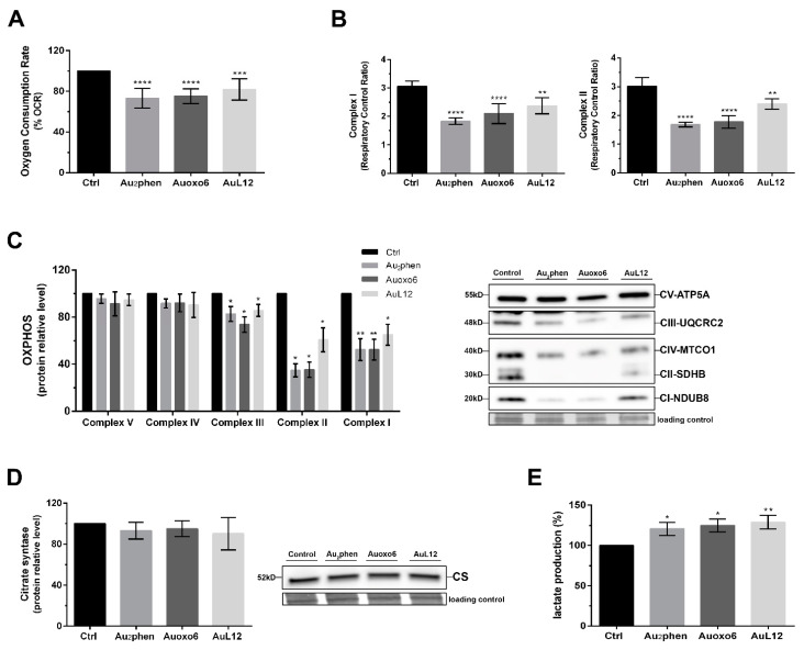
Au2phen ((2,9-dimethyl-1,10-phenanthroline)2Au2(µ-O)2)(PF6)2 and Auoxo6 ((6,6'-dimethyl-2,2'-bipyridine)2Au2(µ-O)2)(PF6)2 are two structurally related gold(III) complexes that were previously reported to display relevant and promising anticancer properties in vitro toward a large number of human cancer cell lines. To expand the knowledge on the molecular mechanisms through which these gold(III) complexes trigger apoptosis in cancer cells, further studies have been performed using A2780 ovarian cancer cells as reference models. For comparative purposes, parallel studies were carried out on the gold(III) complex AuL12 (dibromo(ethylsarcosinedithiocarbamate)gold(III)), whose proapoptotic profile had been earlier characterized in several cancer cell lines. Our results pointed out that all these gold(III) compounds manifest a significant degree of similarity in their cellular and proapoptotic effects; the main observed perturbations consist of potent thioredoxin reductase inhibition, disruption of the cell redox balance, impairment of the mitochondrial membrane potential, and induction of associated metabolic changes. In addition, evidence was gained of the remarkable contribution of ASK1 (apoptosis-signal-regulating kinase-1) and AKT pathways to gold(III)-induced apoptotic signaling. Overall, the observed effects may be traced back to gold(III) reduction and subsequent formation and release of gold(I) species that are able to bind and inhibit several enzymes responsible for the intracellular redox homeostasis, in particular the selenoenzyme thioredoxin reductase.

Keywords: A2780 ovarian cancer cells; apoptosis signal pathway; gold(III)-based compounds; mitochondria; thioredoxin reductase.


Conflict of interest statement

The authors declare no conflict of interest.

Figures

Figure 1
Figure 1
Chemical structure of the investigated gold(III) compounds: Au2phen ((2,9-dimethyl-1,10-phenanthroline)2Au2(µ-O)2)(PF6)2, Auoxo6 ((6,6′-dimethyl-2,2′-bipyridine)2Au2(µ-O)2)(PF6)2 and AuL12 (dibromo(ethylsarcosinedithiocarbamate)gold(III)).
Figure 2
Figure 2
Apoptotic profile analysis and caspases activation: (A) percentages of apoptotic cells shown by flow cytometry analysis of annexin V/propidium iodide-stained A2780 cells treated for 72 h with Au2phen, Auoxo6, and AuL12 IC50 doses; (B) caspase-8, (C) caspase-9, and (D) caspase-3 activity shown by fluorescence-activated cell sorting analysis using FAM FLICA caspase assay kits in A2780 cells treated for 72 h with gold(III) complex concentrations corresponding to their 72 h IC50 dose. Flow cytometric images are representative of three independent experiments. Histograms report the mean values ±SD. The statistical analysis was carried out using one-way ANOVA test followed by Tukey’s multiple comparisons test using GraphPad Prism software v 6.0 (* p < 0.05, ** p < 0.01, *** p < 0.001, **** p < 0.0001).
Figure 3
Figure 3
Effects on thioredoxin reductase activity and cell redox balance. A2780 cells were treated with Au2phen, Auoxo6, and AuL12 72 h IC50 doses: (A) TrxR enzyme inhibition assay was performed after 24 h of treatment using a commercial thioredoxin reductase assay kit, whereby histograms report the percentages of TrxR enzyme activity on gold(III)-treated cells in comparison with controls; (B) intracellular ROS production was evaluated after 48 h of treatment through the detection of fluorogenic H2DCFDA dye using a flow cytometer, whereby histograms report the percentages of fluorescent treated cells compared to controls. Flow cytometric images are representative of three independent experiments. All experiments were performed in triplicate. The statistical analysis was carried out using one-way ANOVA test followed by Tukey’s multiple comparisons test using Graphpad Prism software v 6.0 (* p < 0.05, ** p < 0.01, *** p < 0.001).
Figure 4
Figure 4
Involvement of mitochondrial membrane potential (Δψm) dysfunction in gold(III)-induced apoptosis. A2780 cells were treated for 48 h with Au2phen, Auoxo6, and AuL12 72 h IC50-doses: (A) Δψm values obtained using fluorescent TMRM dye and flow cytometry analysis, whereby flow cytometric images are representative of three independent experiments and histograms report the percentages of treated cells with low TMRM fluorescence compared to controls; (B) Western blot analysis of Bcl-2 and Bax contents, whereby histograms report the normalized mean relative integrated density (±SD) values of Bcl-2 and Bax immunostained bands compared to controls. Representative immunoblots are also shown together with the matching Coomassie-stained PVDF membranes, which were used as loading control. Original Western blot images of Bcl-2 and Bax content are reported in Figure S3. All experiments were performed in triplicate. The statistical analysis was carried out using one-way ANOVA test followed by Tukey’s multiple comparisons test using GraphPad Prism software v 6.0 (* p < 0.05, *** p < 0.001).
Figure 5
Figure 5
Influence of gold(III) complexes on mitochondrial metabolism. A2780 cells were treated for 24 h with Au2phen, Auoxo6, and AuL12 72 h IC50 doses: (A) The percentage of oxygen consumption rate (OCR) and (B) the respiratory control ratio (RCR) values were measured using a Clark-type O2 electrode from Hansatech Instruments (King’s Lynn, Norfolk, UK) as described in Materials and Methods. The protein amount of (C) oxidative phosphorylation (OXPHOS) complexes and (D) citrate synthase (CS) was evaluated using Western blot analysis. Histograms report the normalized mean relative integrated density (±SD) values for each OXPHOS complex, and CS immune-stained bands compared to controls. Representative immunoblots are also shown together with the matching Coomassie-stained PVDF membranes, which were used as loading control. Original Western blot images of OXPHOS and CS content are reported in Figures S4 and S5. (E) The lactate amounts were detected in 1 mL of supernatant medium using a commercial L-lactic acid assay kit (Megazyme, Bray, Ireland). Histograms report the percentages of mean values ±SD with respect to controls. All experiments were performed in triplicate. The statistical analysis was carried out using one-way ANOVA test followed by Tukey’s multiple comparisons test using GraphPad Prism software v 6.0 (* p < 0.05, ** p < 0.01, *** p < 0.001, **** p < 0.0001).
Figure 6
Figure 6
Contributions of ASK1 and AKT in gold(III)-induced apoptotic signaling. A2780 cells were treated for 24 h with Au2phen, Auoxo6, and AuL12 72 h IC50 doses. The phosphorylation levels and total protein amounts of (A) ASK1 and (B) AKT were analyzed through Western blotting. Histograms report the percentages of normalized mean values ± SD of the phospho-ASK1/total ASK1 ratio and phospho-AKT/total AKT ratio compared to controls. The phospho-ASK1/total ASK1 and phospho-AKT/total AKT ratios were obtained from the normalized relative integrated density values of immunostained bands of three independent Western blots. Representative immunoblots are also shown together with the matching Coomassie-stained PVDF membranes, which were used as loading control. Original Western blot images of ASK1 and AKT phosphorylation level and of their total amount are reported in Figures S6 and S7. The statistical analysis was carried out using one-way ANOVA test followed by Tukey’s multiple comparisons test using GraphPad Prism software v 6.0 (* p < 0.05, ** p < 0.01).

References

    1. Nobili S., Mini E., Landini I., Gabbiani C., Casini A., Messori L. Gold Compounds as Anticancer Agents: Chemistry, Cellular Pharmacology, and Preclinical Studies. Med. Res. Rev. 2010;30:550–580. doi: 10.1002/med.20168. - DOI - PubMed
    1. Zou T., Lum C.T., Lok C.-N., Zhang J.-J., Che C.-M. Chemical Biology of Anticancer Gold(III) and Gold(I) Complexes. Chem. Soc. Rev. 2015;44:8786–8801. doi: 10.1039/C5CS00132C. - DOI - PubMed
    1. Casini A., Kelter G., Gabbiani C., Cinellu M.A., Minghetti G., Fregona D., Fiebig H.-H., Messori L. Chemistry, Antiproliferative Properties, Tumor Selectivity, and Molecular Mechanisms of Novel Gold(III) Compounds for Cancer Treatment: A Systematic Study. JBIC J. Biol. Inorg. Chem. 2009;14:1139–1149. doi: 10.1007/s00775-009-0558-9. - DOI - PubMed
    1. Aldinucci D., Ronconi L., Fregona D. Groundbreaking Gold(III) Anticancer Agents. Drug Discov. Today. 2009;14:1075–1076. doi: 10.1016/j.drudis.2009.07.011. - DOI
    1. Bertrand B., Williams M.R.M., Bochmann M. Gold(III) Complexes for Antitumor Applications: An Overview. Chem. Eur. J. 2018;24:11840–11851. doi: 10.1002/chem.201800981. - DOI - PubMed

LinkOut - more resources