. 2021 Aug 9;9(8):982.
doi: 10.3390/biomedicines9080982.

Differentiation Behaviour of Adipose-Derived Stromal Cells (ASCs) Seeded on Polyurethane-Fibrin Scaffolds In Vitro and In Vivo

Affiliations

Differentiation Behaviour of Adipose-Derived Stromal Cells (ASCs) Seeded on Polyurethane-Fibrin Scaffolds In Vitro and In Vivo

Katrin Radeloff et al. Biomedicines. .

Abstract

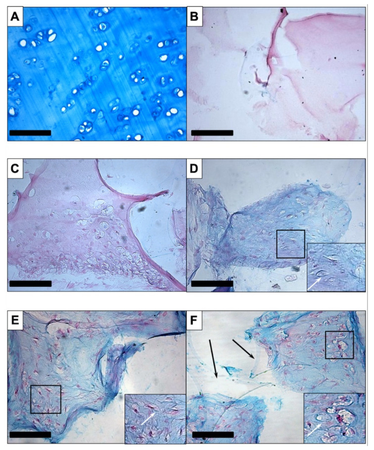
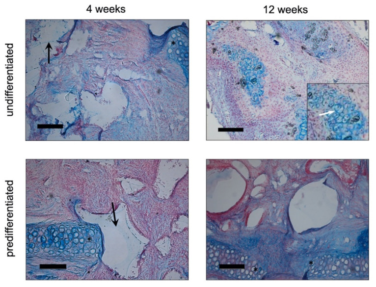
Adipose-derived stromal cells (ASCs) are a promising cell source for tissue engineering and regenerative medicine approaches for cartilage replacement. For chondrogenic differentiation, human (h)ASCs were seeded on three-dimensional polyurethane (PU) fibrin composites and induced with a chondrogenic differentiation medium containing TGF-ß3, BMP-6, and IGF-1 in various combinations. In addition, in vitro predifferentiated cell-seeded constructs were implanted into auricular cartilage defects of New Zealand White Rabbits for 4 and 12 weeks. Histological, immunohistochemical, and RT-PCR analyses were performed on the constructs maintained in vitro to determine extracellular matrix (ECM) deposition and expression of specific cartilage markers. Chondrogenic differentiated constructs showed a uniform distribution of cells and ECM proteins. RT-PCR showed increased gene expression of collagen II, collagen X, and aggrecan and nearly stable expression of SOX-9 and collagen I. Rabbit (r)ASC-seeded PU-fibrin composites implanted in ear cartilage defects of New Zealand White Rabbits showed deposition of ECM with structures resembling cartilage lacunae by Alcian blue staining. However, extracellular calcium deposition became detectable over the course of 12 weeks. RT-PCR showed evidence of endochondral ossification during the time course with the expression of specific marker genes (collagen X and RUNX-2). In conclusion, hASCs show chondrogenic differentiation capacity in vitro with the expression of specific marker genes and deposition of cartilage-specific ECM proteins. After implantation of predifferentiated rASC-seeded PU-fibrin scaffolds into a cartilage defect, the constructs undergo the route of endochondral ossification.

Keywords: ASC; BMP-6; TGF-ß3; adipose-derived stromal cells; chondrogenic differentiation; endochondral ossification; fibrin; polyurethane.


Conflict of interest statement

The authors declare no conflict of interest.

Figures

Figure 1
Figure 1
Procedure of the in vivo studies with New Zealand White Rabbits.
Figure 2
Figure 2
Alcian blue staining: Intense staining was seen in human septal cartilage, the positive control (A). PU-fibrin scaffolds without cells (B) and cell-seeded scaffolds maintained in EM-DMEM (C) revealed no blue staining. Chondrogenic differentiation in polyurethane-fibrin composites was verified by the detection of acid glycosaminoglycans in the extracellular matrix, apparent as extracellular blue-turquoise colouration in the growth factor-treated groups BBT (D), BT (E), and TI (F). Another interesting finding was scattered cartilage lacunae-like structures in the sections of chondrogenic-induced ASC-seeded PU fibrin constructs. Corresponding marked areas were shown enlarged in inserted smaller pictures.The white arrows highlight enhanced staining in the marginal area of the structures. Polygonal, clear sections of the polyurethane foam are highlighted by black arrows in 2F. Magnification is ×200 in all figures; the scale bar represents 100 µm.
Figure 3
Figure 3
Immunohistochemistry with anti-collagen II: An even brown staining of the extracellular matrix of human septal cartilage resulted (A), whereas the negative control, muscular tissue (B), and the cell-seeded PU-fibrin construct maintained in EM-DMEM showed no brown colouration (C). The treated groups revealed weak staining in the BBT-treated group (D) and visible brown staining in the BT (E) and TI (F) group. Magnification is ×200 in all figures; the scale bar represents 100 µm.
Figure 4
Figure 4
Immunohistochemistry with anti-aggrecan: There was clear staining in the septal cartilage (A) and no staining of the muscular tissue (B) or the constructs maintained in EM-DMEM (C). Brown staining was apparent in the treated groups (DF). Magnification is ×200 in all figures; the scale bar represents 100 µm.
Figure 5
Figure 5
RT-PCR analyses of the in vitro PU-fibrin constructs: Relative quantification was performed and is presented as values (∆∆CT values) normalised to the gene expression of the housekeeping gene GAPDH and day 0. SOX-9: No significant differences during the time course and between the different groups. Aggrecan: No significant difference during the time course compared to the control, but a tendency towards increased gene expression in the BBT and BT groups. Collagen II: Significant increase in the BT and TI groups compared to the control after 21 days. Collagen IX: A tendency to increase was also detectable, but no significant difference was evident. Collagen I: After an initial increase compared with the control, no relevant change of gene expression was determined in any group compared to the control group after 21 days. Collagen X: Significant increase in the growth factor groups. Box-Whisker plots show the median, 1st quartile, 3rd quartile as well as minimum and maximum values of ∆∆CT, significance is indicated by asterisks (* p < 0.05, ** p < 0.01, *** p < 0.001, **** p < 0.0001).
Figure 6
Figure 6
Scanning electron microscopy (SEM): The SEM shows that the cell-seeded fibrin hydrogel did not permeate the centre of the polyurethane foam. While the surface of the construct and the pores of the outer part of the PU foam were subtotally covered with the fibrin hydrogel (arrows) ((A,B), which is an enlarged detail of (A)), the centre ((C,D), which is an enlarged detail of (C)) was almost completely empty. Pores of the polyurethane foam are highlighted with asterisks. The scale bar of each pattern represents 100 µm.
Figure 7
Figure 7
Alcian blue staining of cell-seeded PU-fibrin composites in vivo (New Zealand White Rabbit): In the Alcian Blue images, the native auricular cartilage in the region of the defect margin is visible (*). The PU-fibrin construct was slightly thicker than the native cartilage of the auricle, and thus it protruded above the level of the cartilage. Polygonally shaped sections of the PU foam can be seen in the images of the constructs explanted after 4 weeks (black arrows). After 4 weeks, an even distribution of cells and blue-turquoise stained areas could be found within the pores of the PU foam. After 3 months, there were structures with more pronounced blue colouration inside the pores of the construct similar to cartilage lacunae in both groups. The insert shows a clearly accentuated blue staining around these “lacunae” at a higher magnification + highlighted by a white arrow. Qualitatively, no remarkable difference between the groups could be detected. Magnification is × 100 in all figures; the scale bar represents 200 µm.
Figure 8
Figure 8
Von Kossa staining of cell-seeded PU-fibrin composites in vivo (New Zealand White Rabbit): The native auricular cartilage in the region of the defect margin is highlighted with asterisks (*). After 4 weeks, even cell distribution could be seen within the pores of the construct, stained by Nuclear Fast Red. There was no evidence of extracellular calcium deposition. After 12 weeks, distinct black-coloured areas could be detected within the constructs. There was no remarkable difference between both groups. Magnification is ×200 in all figures; the scale bar represents 100 µm.
Figure 9
Figure 9
RT-PCR analysis of cell-seeded PU-fibrin composites in vivo (New Zealand White Rabbit): The gene expression of SOX-9; collagen I, II, and X; and RUNX-2 was analysed and presented as values (∆CT values) normalised to the gene expression of the housekeeping gene GAPDH 4 weeks and 12 weeks after the implantation of the cell-seeded constructs. The chondrogenic marker genes SOX-9 and collagen II were elevated in both groups after 4 and 12 weeks. After implantation, the increase in SOX-9 expression compared to day 0 was only significant for the constructs predifferentiated with BMP-6 and TGF-ß3. Collagen II expression showed a significant increase at 4 and 12 weeks compared to day 0 in the undifferentiated group (EM-DMEM) and at 12 weeks in the predifferentiated group. The expression of the dedifferentiation markers collagen I and collagen X was significantly increased for both groups over the 12 weeks in vivo. RUNX-2, a transcription factor for osteogenic differentiation, was also significantly expressed by both groups during the time course compared to day 0. Box-Whisker plots show the median, 1st quartile, 3rd quartile, and the minimum and maximum values of ∆CT; significance is indicated by asterisks (* p < 0.05, ** p < 0.01, *** p < 0.001, **** p < 0.0001).

References

    1. Frölich K., Kleinsasser N., Rasp G., Staudenmaier R. Navigation-assisted construction of an external ear canal using an autogenous foreskin graft. ORL J. Otorhinolaryngol. Relat. Spec. 2005;67:137–141. doi: 10.1159/000085813. - DOI - PubMed
    1. Giacomini P.G., Ferraro S., Di Girolamo S., Ottaviani F. Large nasal septal perforation repair by closed endoscopically assisted approach. Ann. Plast. Surg. 2011;66:633–636. doi: 10.1097/SAP.0b013e3181dec776. - DOI - PubMed
    1. Detwiller K.Y., Schindler J.S., Schneider D.S., Lindau R. Complex adult laryngotracheal reconstruction with a prefabricated flap: A case series. Head Neck. 2013;3:E376–E380. doi: 10.1002/hed.23335. - DOI - PubMed
    1. Macchiarini P., Walles T., Biancosino C., Mertsching H. First human transplantation of a bioengineered airway tissue. J. Thorac. Cardiovasc. Surg. 2004;128:638–641. doi: 10.1016/j.jtcvs.2004.02.042. - DOI - PubMed
    1. Von Bomhard A., Veit J., Bermueller C., Rotter N., Staudenmaier R., Storck K., The H.N. Prefabrication of 3D cartilage contructs: Towards a tissue engineered auricle—A model tested in rabbits. PLoS ONE. 2013;8:e71667. doi: 10.1371/journal.pone.0071667. - DOI - PMC - PubMed