. 2021 Aug 12;9(8):1003.
doi: 10.3390/biomedicines9081003.

Multiple-Organ Complement Deposition on Vascular Endothelium in COVID-19 Patients

Affiliations

Multiple-Organ Complement Deposition on Vascular Endothelium in COVID-19 Patients

Paolo Macor et al. Biomedicines. .

Abstract

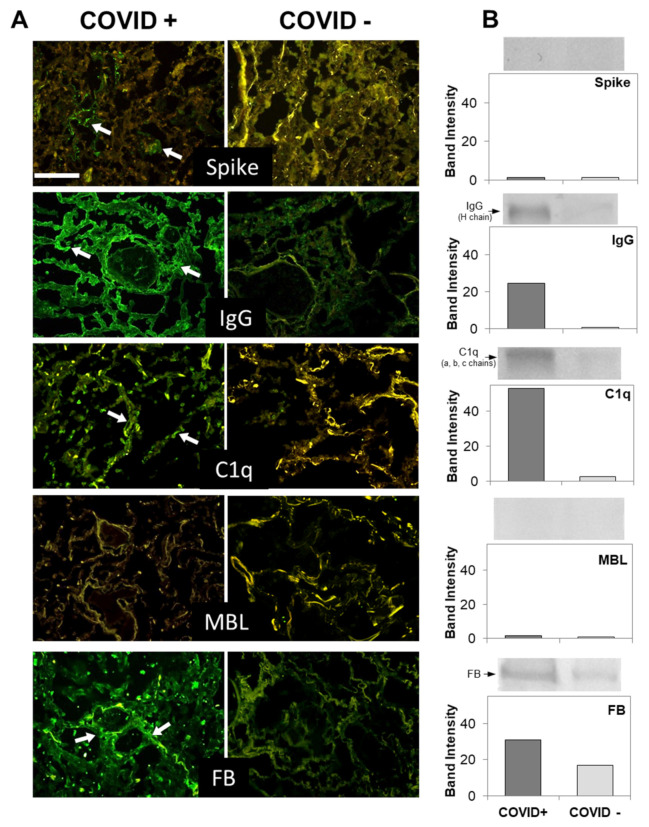
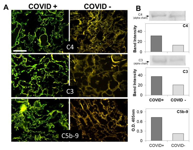
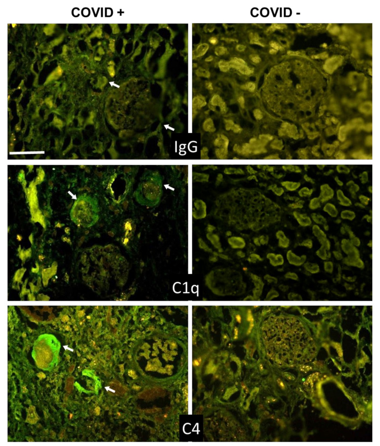
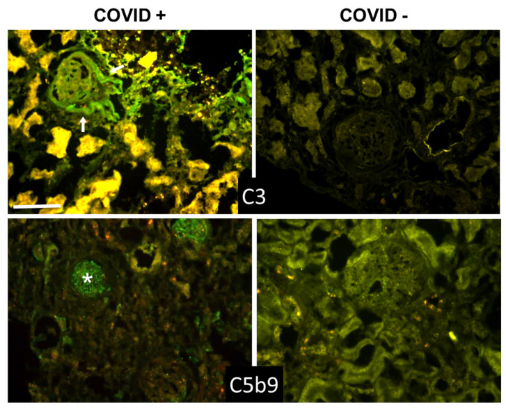
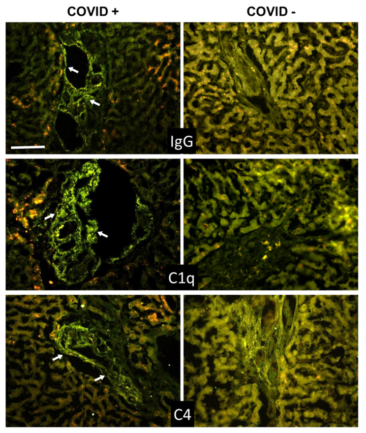
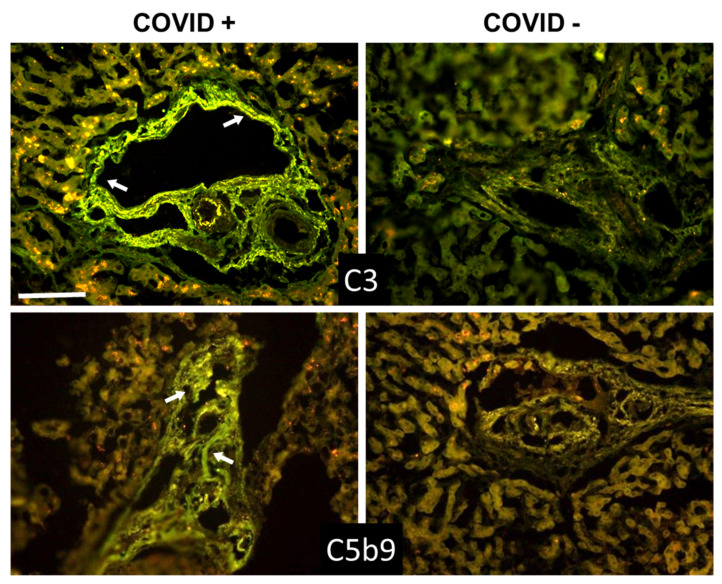
Increased levels of circulating complement activation products have been reported in COVID-19 patients, but only limited information is available on complement involvement at the tissue level. The mechanisms and pathways of local complement activation remain unclear. The aim of this study was to investigate the deposition of complement components in the lungs, kidneys, and liver in patients with COVID-19 patients and to determine the pathway/s of complement activation. We performed immunofluorescence analyses of autopsy specimens of lungs, kidney, and liver from 12 COVID-19 patients who died of acute respiratory failure. Snap-frozen samples embedded in OCT were stained with antibodies against complement components and activation products, IgG, and spike protein of SARS-CoV-2. Lung deposits of C1q, C4, C3, and C5b-9 were localized in the capillaries of the interalveolar septa and on alveolar cells. IgG displayed a similar even distribution, suggesting classical pathway activation. The spike protein is a potential target of IgG, but its uneven distribution suggests that other viral and tissue molecules may be targeted by IgG. FB deposits were also seen in COVID-19 lungs and are consistent with activation of the alternative pathway, whereas MBL and MASP-2 were hardly detectable. Analysis of kidney and liver specimens mirrored findings observed in the lung. Complement deposits were seen on tubules and vessels of the kidney with only mild C5b-9 staining in glomeruli, and on the hepatic artery and portal vein of the liver. Complement deposits in different organs of deceased COVID-19 patients caused by activation of the classical and alternative pathways support the multi-organ nature of the disease and the contribution of the complement system to inflammation and tissue damage.

Keywords: COVID-19; classical pathway; complement activation; multi-organ deposition; spike protein.


Conflict of interest statement

The authors declare that the research was conducted in the absence of any commercial or financial relationships that could be construed as a potential conflict of interest.

Figures

Figure 1
Figure 1
(A) Detection of spike and complement components in lung sections by immunofluorescence analysis. The panel shows representative immunofluorescence images obtained from the analysis of several sections of lung autopsy samples from 12 SARS-CoV-2-positive and 3 negative cases. The sections were stained to reveal the presence of spike protein, IgG, MBL, C1q, and Factor B (FB) (highlighted by a white arrow), as explained in materials and methods, and examined by three independent observers. Scale bar = 50 µm. (B) Evaluation of spike and complement components by western blot in the lungs of a COVID+ and a COVID− patients. The columns represent the staining intensity of the western blot bands quantified using ImageJ (Fuji-NIH). Western blot bands of spike protein, IgG, C1q, MBL and FB are shown on top of the corresponding columns.
Figure 2
Figure 2
(A) Detection of complement components in lung sections by immunofluorescence analysis. The panel shows representative immunofluorescence images obtained from the analysis of several sections of lung autopsy samples from 12 SARS-CoV-2-positive and 3 negative cases. The sections were stained to reveal the presence of C4, C3, and C5b-9, as explained in materials and methods, and examined by three independent observers. Scale bar = 50 µm. (B) Evaluation of complement components by western blot in the lungs of a COVID+ and a COVID− patients. The columns represent the staining intensity of the western blot bands quantified using ImageJ (Fuji-NIH). Western blot bands of C4 and C3 are shown on top of the corresponding columns. C5b-9 levels were measured by ELISA and expressed as OD readings at 405 nm.
Figure 3
Figure 3
Detection of IgG, C1q, and C4 in kidney sections by immunofluorescence analysis. The panel shows representative immunofluorescence images obtained from the analysis of several sections of kidney autopsy samples from 12 SARS-CoV-2-positive and 3 negative cases. See legend to Figure 1 for further details. White arrows show IgG, C1q, and C4 periglomerular deposition. Scale bar = 50 µm.
Figure 4
Figure 4
Detection of C3 and C5b-9 in kidney sections by immunofluorescence analysis. The panel shows representative immunofluorescence images obtained from the analysis of several sections of kidney autopsy samples from 12 SARS-CoV-2-positive and 3 negative cases. See legend to Figure 1 for further details. White arrows in C3 image show periglomerular deposition; * in C5b-9 image shows glomerular staining. Scale bar = 50 µm.
Figure 5
Figure 5
Detection of IgG, C1q, and C4 in liver sections by immunofluorescence analysis. The panel shows representative immunofluorescence images obtained from the analysis of several sections of liver autopsy samples from 12 SARS-CoV-2-positive and 3 SARS-CoV-2-negative cases. See legend to Figure 1 for further details. White arrows indicate vessels in the portal area. Scale bar = 50 µm.
Figure 6
Figure 6
Detection of C3 and C5b-9 in liver sections by immunofluorescence analysis. The panel shows representative immunofluorescence images obtained from the analysis of several sections of liver autopsy samples from 12 SARS-CoV-2-positive and 3 SARS-CoV-2-negative cases. See legend to Figure 1 for further details. White arrows indicate vessels in the portal area. Scale bar = 50 µm.

Update of

References

    1. Remuzzi A., Remuzzi G. COVID-19 and Italy: What next? Lancet. 2020;395:1225–1228. doi: 10.1016/S0140-6736(20)30627-9. - DOI - PMC - PubMed
    1. Fu L., Wang B., Yuan T., Chen X., Ao Y., Fitzpatrick T., Li P., Zhou Y., Lin Y.F., Duan Q., et al. Clinical characteristics of coronavirus disease 2019 (COVID-19) in China: A systematic review and meta-analysis. J. Infect. 2020;80:656–665. doi: 10.1016/j.jinf.2020.03.041. - DOI - PMC - PubMed
    1. Schaefer I.M., Padera R.F., Solomon I.H., Kanjilal S., Hammer M.M., Hornick J.L., Sholl L.M. In situ detection of SARS-CoV-2 in lungs and airways of patients with COVID-19. Mod. Pathol. 2020;33:2104–2114. doi: 10.1038/s41379-020-0595-z. - DOI - PMC - PubMed
    1. Li P., Xie M., Zhang W. Clinical characteristics and intrauterine vertical transmission potential of coronavirus disease 2019 infection in 9 pregnant women: A retrospective review of medical records. Am. J. Obstet. Gynecol. 2020;223:955–956. doi: 10.1016/j.ajog.2020.08.059. - DOI - PMC - PubMed
    1. Hamming I., Timens W., Bulthuis M.L., Lely A.T., Navis G., van Goor H. Tissue distribution of ACE2 protein, the functional receptor for SARS coronavirus. A first step in understanding SARS pathogenesis. J. Pathol. 2004;203:631–637. doi: 10.1002/path.1570. - DOI - PMC - PubMed

LinkOut - more resources