Review
. 2021 Aug 14;9(8):1014.
doi: 10.3390/biomedicines9081014.

Extra- and Intra-Cellular Mechanisms of Hepatic Stellate Cell Activation

Affiliations
Review

Extra- and Intra-Cellular Mechanisms of Hepatic Stellate Cell Activation

Yufei Yan et al. Biomedicines. .

Abstract

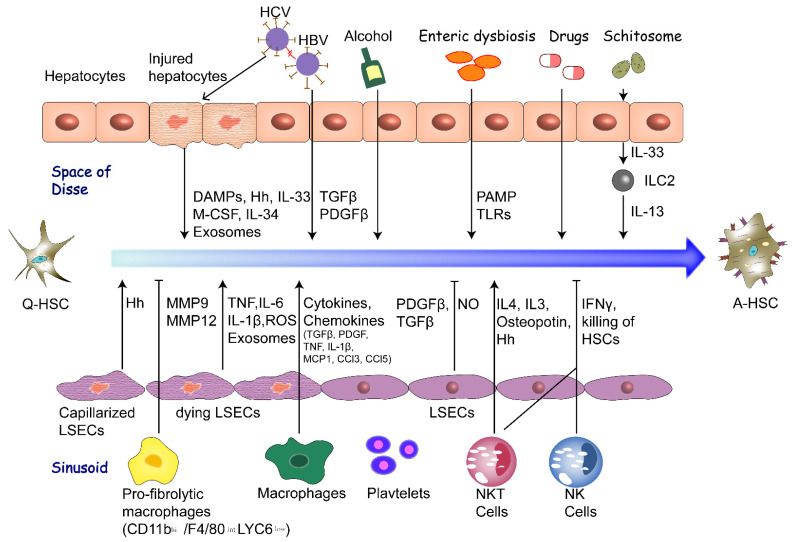
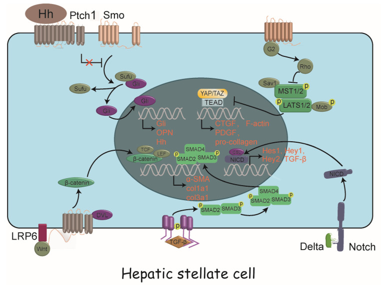
Hepatic fibrosis is characterized by the pathological accumulation of extracellular matrix (ECM) in the liver resulting from the persistent liver injury and wound-healing reaction induced by various insults. Although hepatic fibrosis is considered reversible after eliminating the cause of injury, chronic injury left unchecked can progress to cirrhosis and liver cancer. A better understanding of the cellular and molecular mechanisms controlling the fibrotic response is needed to develop novel clinical strategies. It is well documented that activated hepatic stellate cells (HSCs) is the most principal cellular players promoting synthesis and deposition of ECM components. In the current review, we discuss pathways of HSC activation, emphasizing emerging extra- and intra-cellular signals that drive this important cellular response to hepatic fibrosis. A number of cell types and external stimuli converge upon HSCs to promote their activation, including hepatocytes, liver sinusoidal endothelial cells, macrophages, cytokines, altered ECM, hepatitis viral infection, enteric dysbiosis, lipid metabolism disorder, exosomes, microRNAs, alcohol, drugs and parasites. We also discuss the emerging signaling pathways and intracellular events that individually or synergistically drive HSC activation, including TGFβ/Smad, Notch, Wnt/β-catenin, Hedgehog and Hippo signaling pathways. These findings will provide novel potential therapeutic targets to arrest or reverse fibrosis and cirrhosis.

Keywords: hepatic fibrosis; hepatic stellate cell; myofibroblast; signal pathway.


Conflict of interest statement

The authors have declared that no conflict of interest exists.

Figures

Figure 1
Figure 1
Extracellular factors promoting HSC activation. Extracellular factors including stimulation of various cell types, cyto-kines, altered ECM, hepatitis viral infection, enteric dysbiosis, lipid metabolism disorder, exosomes, alcohol, drugs (MTX, APAP) and schistosome promote or inhibit the activation of HSCs through production of various cytokines and other signaling molecules. The characteristics of A-HSCs include proliferation, contractility, fibrogenesis, inflammatory sig-naling, loss of retinoid and enhanced ECM production. HSC, hepatic stellate cell; DAMPs, damage-associated molecular patterns; Hh, Hedgehog signaling; IL-33, interleukin-33; M-CSF, macrophage colony-stimulating factor; TGFβ, Transforming growth factor β; PDGFβ, Platelet derived growth factor β; PAMP, pathogen-associated molecular patterns; TLRs, Toll-like receptors; MMP, matrix metalloproteinases; TNF, tumor necrosis factor; ROS, reactive oxygen species; MCP1, monocyte chemo-tactic protein-1; NO, nitric oxide; IFNγ, interferon γ; NK, natural killer; NKT, natural killer T; MTX, methotrexate; APAP, acetaminophen; hi, high expression; low, low expression; int, intermediate expression.
Figure 2
Figure 2
Intracellular signaling pathways driving HSC activation. A panoply of signals drive HSC activation, including TGF-β/SMAD pathway, Notch signaling, Wnt/β-catenin signaling, Hedgehog signaling and Hippo signaling, with complex crosstalk between them. HSC, hepatic stellate cell; Hh, hedgehog; Ptch1, patched 1; Smo, smoothened; Sufu, suppressor of fused; Gli, glioma-associated oncogene homolog; OPN, osteopontin; TCF, T lymphocyte factor; LEF, lymphocyte enhancer factor; LRP6, low-density lipoprotein receptor 6; DVL, disheveled; SMAD, small mother against decapentaplegic; α-SMA, α-smooth muscle actin; col1a1, collagen I α-1; TGF-β,transforming growth factor β; Sav1, Salvador family WW domain containing protein 1; MST1/2, mammalian STE20-like protein kinase 1 and 2; LATS1/2, large tumor suppressor kinase 1 and 2; Mob, monopolar spindle-one-binder protein; YAP, yes-associated protein; TAZ, transcriptional coactivator with PDZ-binding motif; TEAD, TEA domain transcription factor; CTGF, connective tissue growth factor; PDGF, platelet derived growth factor; NICD, Notch intracellular domain; CSL, CBF-1, Suppressor of hairless, Lag-2; Hes, hairy/enhancer of split ; Hey, hairy/enhancer of split related with YRPW motif.

References

    1. Torres-Hernandez A., Wang W., Nikiforov Y., Tejada K., Torres L., Kalabin A., Wu Y., Haq M.I.U., Khan M.Y., Zhao Z., et al. Targeting SYK signaling in myeloid cells protects against liver fibrosis and hepatocarcinogenesis. Oncogene. 2019;38:4512–4526. doi: 10.1038/s41388-019-0734-5. - DOI - PubMed
    1. Lee U.E., Friedman S.L. Mechanisms of hepatic fibrogenesis. Best Pract. Res. Clin. Gastroenterol. 2011;25:195–206. doi: 10.1016/j.bpg.2011.02.005. - DOI - PMC - PubMed
    1. Parola M., Pinzani M. Liver fibrosis: Pathophysiology, pathogenetic targets and clinical issues. Mol. Asp. Med. 2019;65:37–55. doi: 10.1016/j.mam.2018.09.002. - DOI - PubMed
    1. Asrani S.K., Devarbhavi H., Eaton J., Kamath P.S. Burden of liver diseases in the world. J. Hepatol. 2019;70:151–171. doi: 10.1016/j.jhep.2018.09.014. - DOI - PubMed
    1. Qi X., De Stafano V., Guo X. Treatment of Patients with Cirrhosis. N. Engl. J. Med. 2016;375:2103–2104. doi: 10.1056/NEJMc1612334. - DOI - PubMed

LinkOut - more resources