Review
. 2021 Jul 29;12(8):1180.
doi: 10.3390/genes12081180.

Intermixing the OPN1LW and OPN1MW Genes Disrupts the Exonic Splicing Code Causing an Array of Vision Disorders

Affiliations
Review

Intermixing the OPN1LW and OPN1MW Genes Disrupts the Exonic Splicing Code Causing an Array of Vision Disorders

Maureen Neitz et al. Genes (Basel). .

Abstract

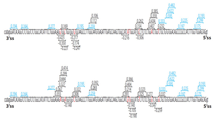
Light absorption by photopigment molecules expressed in the photoreceptors in the retina is the first step in seeing. Two types of photoreceptors in the human retina are responsible for image formation: rods, and cones. Except at very low light levels when rods are active, all vision is based on cones. Cones mediate high acuity vision and color vision. Furthermore, they are critically important in the visual feedback mechanism that regulates refractive development of the eye during childhood. The human retina contains a mosaic of three cone types, short-wavelength (S), long-wavelength (L), and middle-wavelength (M) sensitive; however, the vast majority (~94%) are L and M cones. The OPN1LW and OPN1MW genes, located on the X-chromosome at Xq28, encode the protein component of the light-sensitive photopigments expressed in the L and M cones. Diverse haplotypes of exon 3 of the OPN1LW and OPN1MW genes arose thru unequal recombination mechanisms that have intermixed the genes. A subset of the haplotypes causes exon 3- skipping during pre-messenger RNA splicing and are associated with vision disorders. Here, we review the mechanism by which splicing defects in these genes cause vision disorders.

Keywords: X-linked cone dysfunction; color vision; colorblindness; cone photopigment; exon skipping; myopia.


Conflict of interest statement

M. Neitz and J. Neitz are co-founders of SightGlass Vision and inventors of the eyeglasses currently being evaluated in NCT NCT03623074. They and the University of Washington receive licensing fees and royalties from patents related to myopia.

Figures

Figure 1
Figure 1
Opsin minigene splicing assay. Top: The OPN1LW minigene is OPN1LW cDNA with introns 2 and 3 inserted. The introns are labeled, and the numbered red rectangles are the exons that were identical in sequence for all minigenes. The gray rectangle is exon 3, which varied in sequence among the minigenes. The effects of different SNP combinations in exon 3 on splicing were measured by first transfecting HEK293 cells with minigenes. mRNA was isolated from HEK293 cells, and the amount of correctly spliced mRNA was quantified. Data in Table 2 are from reference [22], and the percentage of correctly spliced mRNA was measured using capillary gel electrophoresis.
Figure 2
Figure 2
Opsin gene array structure, cone mosaic, and color vision phenotype. The numbered rectangles represent the exons of the OPN1LW and OPN1MW genes. Black rectangles represent exons 1 and 6. Exons 2, 3, and 4 are polymorphic among and between the OPN1LW and OPN1MW genes and thus are colored yellow. Exon 5 encodes two amino acid differences that functionally distinguish L from M photopigments [34]; therefore, red rectangles represent exon 5 of OPN1LW genes, and green rectangles represent exon 5 of OPN1LW genes. (A) An opsin gene array with the two expressed positions occupied by OPN1LW and OPN1MW genes will give rise to a retinal cone mosaic with L cones (red circles), M cones (green circles), and S cones (blue circles) and will confer normal trichromatic color vision. (B) An opsin gene array with only one OPN1LW gene (second and third genes N = 0) or the two expressed positions occupied by OPN1LW genes (second gene N = 1) gives rise to a retinal mosaic with L cones and S cones, but no M cones, and will underlie deutan CVD. (C) An opsin gene array with one OPN1MW gene (second and third genes N = 0) or with both expressed positions (second gene N = 1) occupied by an OPN1MW gene will give rise to a retinal mosaic with only S cones and M cones, but no L cones and will underlie protan CVD. Please see references [8,69] for recent reviews of the genetics of normal and defective color vision.
Figure 3
Figure 3
(A) Arrays with an OPN1LW gene followed by an OPN1MW gene, each with haplotypes that splice normally, give rise to L and M cones that have a normal amount of photopigment, which in turn confers normal, trichromatic red-green color vision. Exons are represented by the numbered rectangles, and the color-coding is the same as that described in Figure 2. (B) An array with a single OPN1LWLIAVA gene does not produce any functional photopigment, so all cones except-S cones are devoid of pigment, and a male with this array is an obligate Blue Cone Monochromacy (BCM). (C) An array in which the first two genes have the LIAVA haplotype will cause BCM regardless of whether the second gene encodes an OPN1LW or OPN1MW gene (indicated by a yellow rectangle for exon 5) because all cones except S cones will be devoid of photopigment. (D) An array in which the first gene is OPN1LWLIAVA and the second gene is either OPN1MWMVVVA or OPN1MWMIAVA will cause protanopia in males because the L cones will be devoid of photopigment, and the M cones will have photopigment. Thus, color vision will be mediated by M and S cones. (E) An array in which the OPN1LW gene has a normal (non-exon 3-skipping) haplotype and the second gene is an OPN1MWLIAVA will cause deuteranopia in males because only L cones and S cones will contain functional photopigment. Arrays in this figure correspond to those found in patients described in Tables S1–S3 [22,27,31,33,39].
Figure 4
Figure 4
Opsin gene arrays with LVAVA haplotype and the associated color vision. Rectangles represent exons; see Figure 2 and Figure 3 for a description of the color-coding. For example, the yellow rectangle representing exon 5 indicates the gene may be OPN1LW or OPN1MW. (A) Array with a single opsin gene that is either OPN1LWLVAVA or OPN1MWLVAVA. A male with an array like this will be an obligate deuteranope if the gene is OPN1LW or an obligate protanope if the gene is OPN1MW. Deuteranopes have functional S and L cones; protanopes have function S and M cones. (B) An array in which both expressed positions have the LVAVA haplotype. In a male with this array all non-S cones will have a small amount of functional photopigment. The color vision phenotype depends on whether the second gene is L (deutan) or M (normal). (C,D) An array with OPN1LWLVAVA in the first position and an OPN1LW or OPN1MW gene with the MVVVA (C) or LVAIA (D) haplotype in the second position. Males with one of these arrays will have CVD or normal color vision depending on whether the second gene is OPN1LW (deutan) or OPN1MW (normal). (E) An array with OPN1LWLVAVA and OPN1MWMVAVA. A male with this array will have functional L and M cones and thus normal color vision. Arrays in this figure are based on patients described in Tables S4–S6 [22,31,32,33,38,39,81].
Figure 5
Figure 5
Map showing quantitative effects of hexamers as exonic splicing elements. The upper and lower sequences are for OPN1LW/OPN1MW exon 3, including the locations of the 5′ and 3′ splice sites (5′ss and 3′ss, respectively). The letters in red font show the common SNPs at each of the eight exon 3 SNPs and, from left to right, correspond to cDNA positions c.453, c.457, c.465, c.511, c.513, c.521, c.528, and c.532. The numbers in blue indicate hexamers that are not affected by the SNPs; the numbers in black change depending on the variant SNPs (upper vs. lower map). Positive values are ESEs; negative values are ESSs. The larger the absolute value of the number, the greater the effect on splicing. The values indicating the impact of each hexamer on splicing are from reference [82].

References

    1. Cartegni L., Chew S.L., Krainer A.R. Listening to silence and understanding nonsense: Exonic mutations that affect splicing. Nat. Rev. Genet. 2002;3:285–298. doi: 10.1038/nrg775. - DOI - PubMed
    1. Pagani F., Stuani C., Tzetis M., Kanavakis E., Efthymiadou A., Doudounakis S., Casals T., Baralle F.E. New type of disease causing mutations: The example of the composite exonic regulatory elements of splicing in CFTR exon 12. Hum. Mol. Genet. 2003;12:1111–1120. doi: 10.1093/hmg/ddg131. - DOI - PubMed
    1. Wang G.S., Cooper T.A. Splicing in disease: Disruption of the splicing code and the decoding machinery. Nat. Rev. Genet. 2007;8:749–761. doi: 10.1038/nrg2164. - DOI - PubMed
    1. Ward A.J., Cooper T.A. The pathobiology of splicing. J. Pathol. 2010;220:152–163. doi: 10.1002/path.2649. - DOI - PMC - PubMed
    1. Sterne-Weiler T., Howard J., Mort M., Cooper D.N., Sanford J.R. Loss of exon identity is a common mechanism of human inherited disease. Genome Res. 2011;21:1563–1571. doi: 10.1101/gr.118638.110. - DOI - PMC - PubMed

Publication types

LinkOut - more resources