Review
. 2021 Aug 13;12(8):1241.
doi: 10.3390/genes12081241.

An Overview of the Genetics of ABCA4 Retinopathies, an Evolving Story

Affiliations
Review

An Overview of the Genetics of ABCA4 Retinopathies, an Evolving Story

Saoud Al-Khuzaei et al. Genes (Basel). .

Abstract

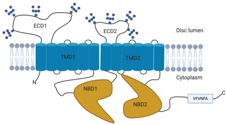
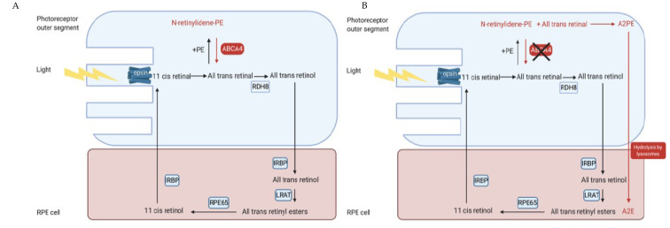
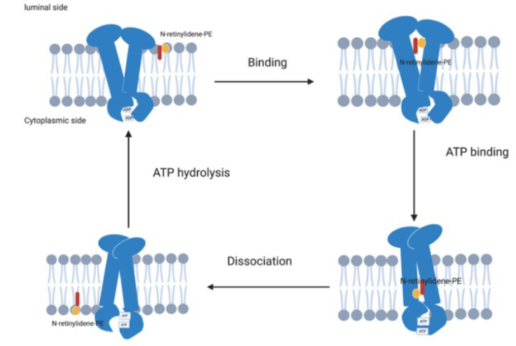
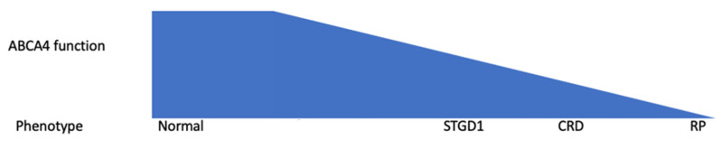
Stargardt disease (STGD1) and ABCA4 retinopathies (ABCA4R) are caused by pathogenic variants in the ABCA4 gene inherited in an autosomal recessive manner. The gene encodes an importer flippase protein that prevents the build-up of vitamin A derivatives that are toxic to the RPE. Diagnosing ABCA4R is complex due to its phenotypic variability and the presence of other inherited retinal dystrophy phenocopies. ABCA4 is a large gene, comprising 50 exons; to date > 2000 variants have been described. These include missense, nonsense, splicing, structural, and deep intronic variants. Missense variants account for the majority of variants in ABCA4. However, in a significant proportion of patients with an ABCA4R phenotype, a second variant in ABCA4 is not identified. This could be due to the presence of yet unknown variants, or hypomorphic alleles being incorrectly classified as benign, or the possibility that the disease is caused by a variant in another gene. This underlines the importance of accurate genetic testing. The pathogenicity of novel variants can be predicted using in silico programs, but these rely on databases that are not ethnically diverse, thus highlighting the need for studies in differing populations. Functional studies in vitro are useful towards assessing protein function but do not directly measure the flippase activity. Obtaining an accurate molecular diagnosis is becoming increasingly more important as targeted therapeutic options become available; these include pharmacological, gene-based, and cell replacement-based therapies. The aim of this review is to provide an update on the current status of genotyping in ABCA4 and the status of the therapeutic approaches being investigated.

Keywords: ABCA4; ABCA4-associated retinopathies; Stargardt disease; genetic testing; phenocopies.


Conflict of interest statement

The authors declare no conflict of interest. The funders had no role in the design of the study; in the collection, analyses, or interpretation of data; in the writing of the manuscript, or in the decision to publish the results.

Figures

Figure 1
Figure 1
Colour fundus photographs: (A) foveal atrophy surrounded by a minimal amount of flecks; and (B) widespread flecks that would be consistent with a fundus flavimaculatus phenotype.
Figure 2
Figure 2
Schematic diagram of the structure of the ABCA4 protein showing the two transmembrane domains (TMD), the nucleotide-binding domains (NBD), and exocytoplasmic domains (ECD) that contain N-linked oligosaccharide chains and the C-terminal VFVNFA motif. Adapted from [33,37,38]. Created with BioRender.com (accessed on 1 June 2021).
Figure 3
Figure 3
Schematic diagram illustrating the visual cycle in the photoreceptor outer segments and the RPE. (A) Light photobleaches the opsin and isomerises the 11-cis-retinal to ATR. Some ATR reversibly reacts with PE to form NrPE, which is flipped onto the cytoplasmic side by the ABCA4 protein. The NrPE is then hydrolysed to PE and ATR, thus preventing the accumulation of ATR on the luminal side. The ATR is then reduced to all-trans-retinol by RDH8 and then transported to the RPE cell by IRBP. In the RPE, the all-trans-retinol is esterified to all-trans-retinyl esters by LRAT, which is then converted to 11-cis-retinol by RPE65 isomerohydrolase and then oxidized to 11-cis-retinal by RDH and transported back to the photoreceptors by IRBP. (B) Schematic diagram illustrating the visual cycle in the presence of ABCA4 dysfunction. Dysfunction of the ABCA4 protein prevents the flipping of the NrPE from the luminal side to the cytoplasmic side of the photoreceptor outer segments, meaning that the NrPE accumulates and condenses with all-trans-retinal into A2PE. The photoreceptor outer segments are then shed and phagocytosed by the RPE cell, which then hydrolyse the A2PE to A2E [56]. Created with BioRender.com (accessed on 1 June 2021). Abbreviations: ATR: All-trans-retinal; A2E: N-retinyl-N-retinylidene ethanolamine; A2PE phosphatidyl-pyridinium bisretinoid; IRBP: inter photoreceptor binding protein; LRAT: lecithin retinol acyltransferase, NrPE: N-retinylidene phosphatidylethanolamine; PE: phosphatidylethanolamine; RDH8: retinol dehydrogenase 8; RPE: retinal pigment epithelial.
Figure 4
Figure 4
Schematic diagram of the ABCA4 protein actively transporting NrPE from the luminal side of the photoreceptor disc membrane to the cytoplasmic side. The ADP is initially bound to the NBD and the ABCA4 binds the NrPE on the luminal side of the photoreceptor disc membrane; this is followed by binding of ATP to the NBDs, leading to a conformational change that creates a low affinity binding site on the cytoplasmic side, resulting in dissociation of the NrPE from the ABCA4 protein and followed by hydrolysis of the ATP, returning the ABCA4 to its primary conformation. Adapted from Molday et al. [37]. Created with BioRender.com (accessed on 1 June 2021). Abbreviations: NBD: nuclear binding domain; NrPE: N-retinylidene phosphatidylethanolamine.
Figure 5
Figure 5
Schematic diagram illustrating the correlation between the amount of functional ABCA4 protein and the phenotype based on early studies that predicted that a lower amount of functioning ABCA4 protein was associated with more severe phenotypes. Figure adapted from Maugeri et al. [53].

References

    1. Blacharski P. Retinal Dystrophies and Degenerations. Raven Press; New York, NY, USA: 1988. pp. 135–159.
    1. Allikmets R., Singh N., Sun H., Shroyer N.F., Hutchinson A., Chidambaram A., Gerrard B., Baird L., Stauffer D., Peiffer A., et al. A photoreceptor cell-specific ATP-binding transporter gene (ABCR) is mutated in recessive Starqardt macular dystrophy. Nat. Genet. 1997;15:236–246. doi: 10.1038/ng0397-236. - DOI - PubMed
    1. Yatsenko A.N., Shroyer N.F., Lewis R.A., Lupski J.R. Late-onset Stargardt disease is associated with missense mutations that map outside known functional regions of ABCR (ABCA4) Hum. Genet. 2001;108:346–355. doi: 10.1007/s004390100493. - DOI - PubMed
    1. Jaakson K., Zernant J., Külm M., Hutchinson A., Tonisson N., Glavac D., Ravnik-Glavac M., Hawlina M., Meltzer M.R., Caruso R.C., et al. Genotyping microarray (gene chip) for the ABCR (ABCA4) gene. Hum. Mutat. 2003;22:395–403. doi: 10.1002/humu.10263. - DOI - PubMed
    1. Stargardt K. Über familiäre, progressive Degeneration in der Maculagegend des Auges. Albrecht Graefes Arch. Ophthalmol. 1909;71:534–550. doi: 10.1007/BF01961301. - DOI

Publication types

MeSH terms

Substances