. 2021 Jul 21;11(8):723.
doi: 10.3390/life11080723.

Intracellular Development of Resident Cardiac Stem Cells: An Overlooked Phenomenon in Myocardial Self-Renewal and Regeneration

Affiliations

Intracellular Development of Resident Cardiac Stem Cells: An Overlooked Phenomenon in Myocardial Self-Renewal and Regeneration

Galina Belostotskaya et al. Life (Basel). .

Abstract

At present, the approaches aimed at increasing myocardial regeneration after infarction are not available. The key question is the identity of cells capable of producing functional cardiac myocytes (CMs), replenishing those lost during ischemia. With identification of resident cardiac stem cells (CSCs), it has been supposed that this cell population may be crucial for myocardial self-renewal and regeneration. In the last few years, the focus has been shifted towards another concept, implying that new CMs are produced by dedifferentiation and proliferation of mature CMs. The observation that CSCs can undergo development inside immature cardiac cells by formation of "cell-in-cell structures" (CICSs) has enabled us to conclude that encapsulated CICSs are implicated in mammalian cardiomyogenesis over the entire lifespan. Earlier we demonstrated that new CMs are produced through formation of CSC-derived transitory amplifying cells (TACs) either in the CM colonies or inside encapsulated CICSs. In this study, we described the phenomenon of CSC penetration into mature CMs, resulting in the formation of vacuole-like CICSs (or non-encapsulated CICSs) containing proliferating CSCs with subsequent differentiation of CSC progeny into TACs and their release. In addition, we compared the phenotypes of TACs derived from encapsulated and non-encapsulated CICSs developing in immature and mature CMs, respectively.

Keywords: cardiac stem cells (CSCs); cardiomyocytes (CMs); cardiomyogenesis; myocardial ischemia; regeneration; transitory amplifying cells (TACs); “cell-in-cell structures” (CICSs).


Conflict of interest statement

The authors declare no conflict of interest.

Figures

Figure 1
Figure 1
Cellular structures, identified in the suspension of the myocardial cells in adult rats two weeks after permanent coronary occlusion in the infarct zone (AC), peri-infarct area (DF), and after 40-min ischemia/reperfusion (GI). Confocal and fluorescent microscopy: c-kit+-, Sca-1+-, and Islet-1+- CSCs (green); α-sarcomeric actinin (red); and nuclei (Hoechst 33342, blue).
Figure 2
Figure 2
The average number of encapsulated CICSs in infarct, peri-infarct, and intact areas of the heart in experimental groups two weeks after surgery in the Control (a) 2 weeks after permanent myocardial infarction (b) and 40-min ischemia/reperfusion (c). The data are mean ± standard deviation. SHAM—sham-operated animals, PCAO—permanent coronary artery occlusion, MIRI—myocardial ischemia for 40 min with reperfusion; * p < 0.05 versus the respective value in SHAM group.
Figure 3
Figure 3
Variants of cellular structures revealed in the suspension of the myocardial cells in adult rats from the infarct area at different time points after permanent coronary occlusion. (A) Myocardial fragment with mature CMs and TACs; (B) accumulation of undifferentiated TACs, descendants of c-kit+- CSCs; (C) accumulation of cardio-positive TACs; (D) colony of fibroblasts (at the bottom) and dead CM (at the top); (E) immature c-kit+- CICS (at the top) and capsule of opened/ruptured CICS (at the bottom); (F) small colony of undifferentiated c-kit+- CSC (at the top) and large differentiated colony c-kit+- CSC (at the bottom). Confocal and fluorescent microscopy: c-kit+- CSCs (green), α-sarcomeric actinin (red), and nuclei (Hoechst 3342, blue).
Figure 4
Figure 4
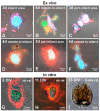
Variants of non-encapsulated CICSs in the suspension of the myocardial cells in adult rats after permanent coronary occlusion (ex vivo) and in the culture of the myocardial cells in rats (in vitro). Ex vivo: Cellular structures from the infarct zone (A), peri-infarct area (C,E) and the area remote from the infarct zone (B,D,F), demonstrating intracellular development of CSCs with formation of non-encapsulated CICSs. A—free vacuole with TACs of c-kit+- type; B—disintegrating CM, releasing 2 vacuoles with c-kit+- TACs of various inner maturity; C—3 vacuoles with c-kit+- TACs inside, located in the cytoplasm of one CM; D, E—vacuoles with c-kit+- TACs inside mature CMs; F—mature CM with abundant c-kit+- TACs inside. In vitro: non-encapsulated CICSs in newborn rats on day 5 in vitro (DIV) –with c-kit+ CSCs and rhodamine phalloidin (G) and on DIV 11 with Islet-1+ CSCs and cardiac myosin (H). Non-encapsulated CICSs in 8-day old rat on DIV 13 (light microscopy—(I)). Confocal and fluorescent microscopy: c-kit+- and Islet-1+- CSCs (green), α-sarcomeric actinin, rhodamine phalloidin and cardiac myosin (red), nuclei (Hoechst 33342, blue).
Figure 5
Figure 5
Confocal microscopy of cellular structures revealed when culturing the myocardial cells of variously aged rats. Newborn rats: on day 5 in vitro (DIV)—(A); on DIV 10—(D); on DIV 15—(G) and on DIV 11—(J). Twenty-day-old rats on DIV 11—(B); on DIV 5 (E,K); and on DIV 7—(H). Forty-day-old rats on DIV 22—(C,F,I) and on DIV 6 (L). Confocal microscopy using proliferation marker Ki67 (green); rhodamine phalloidin (red); of antibodies to Islet-1, c-kit, and Sca-1 CSCs (green); sarcomeric actin (red), and nuclei (Hoechst 33342, blue).
Figure 6
Figure 6
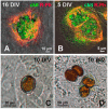
Variants of CICSs in the culture of the neonatal myocardium cells of rats (in vitro). (A,B) confocal microscopy; (C,D) light microscopy (x40).Upper row: (A) non-encapsulated CICS on day 16 in vitro (DIV), formed with CSC of c-kit+ type; (B) non-encapsulated CICS on DIV 5 with two c-kit+- vacuoles inside. Bottom row: (C) encapsulated CICS; (D) 2 encapsulated CICSs with two c-kit+- capsules inside each. Confocal microscopy using antibodies to c-kit CSCs (green) and rhodamine phalloidin (red).
Figure 7
Figure 7
Cellular structures in the infarct area (AC,FI,K), peri-infarct area (D), and the area remote from the infarct (E,J,L). (A,B,L) Encapsulated CICSs. (CE,J,K) Non-encapsulated CICSs. (A,L) (bottom) Release of TACs from encapsulated CICSs. (GI) Mass egress of TACs from mature CMs. (F) Population of TACs inside the mucin mass after exiting CICS. Confocal and fluorescent microscopy with staining: c-kit+- CSCs (green), α-sarcomeric actinin (red), and nuclei (Hoechst 33342, blue).

References

    1. Beltrami A.P., Barlucchi L., Torella D., Baker M., Limana F., Chimenti S., Kasahara H., Rota M., Musso E., Urbanek K., et al. Adult Cardiac Stem Cells Are Multipotent and Support Myocardial Regeneration. Cell. 2003;114:763–776. doi: 10.1016/S0092-8674(03)00687-1. - DOI - PubMed
    1. Cai C.-L., Liang X., Shi Y., Chu P.-H., Pfaff S.L., Chen J., Evans S. Isl1 Identifies a Cardiac Progenitor Population that Proliferates Prior to Differentiation and Contributes a Majority of Cells to the Heart. Dev. Cell. 2003;5:877–889. doi: 10.1016/S1534-5807(03)00363-0. - DOI - PMC - PubMed
    1. Parmacek M., Epstein J.A. Pursuing Cardiac Progenitors: Regeneration Redux. Cell. 2005;120:295–298. doi: 10.1016/j.cell.2005.01.025. - DOI - PubMed
    1. Anversa P., Kajstura J., Leri A., Bolli R. Life and Death of Cardiac Stem Cells: A Paradigm Shift in Cardiac Biology. Circulation. 2006;113:1451–1463. doi: 10.1161/CIRCULATIONAHA.105.595181. - DOI - PubMed
    1. Choi Y.-H., Saric T., Nasseri B., Hühn S., van Linthout S., Hetzer R., Tschöpe C., Stamm C. Cardiac Cell Therapies: The Next Generation. Cardiovasc. Ther. 2010;29:2–16. doi: 10.1111/j.1755-5922.2010.00191.x. - DOI - PubMed

LinkOut - more resources