. 2021 Jul 27;10(8):1900.
doi: 10.3390/cells10081900.

Multisystemic Cellular Tropism of SARS-CoV-2 in Autopsies of COVID-19 Patients

Affiliations

Multisystemic Cellular Tropism of SARS-CoV-2 in Autopsies of COVID-19 Patients

Dickson W L Wong et al. Cells. .

Abstract

Multiorgan tropism of SARS-CoV-2 has previously been shown for several major organs. We have comprehensively analyzed 25 different formalin-fixed paraffin-embedded (FFPE) tissues/organs from autopsies of fatal COVID-19 cases (n = 8), using histopathological assessment, detection of SARS-CoV-2 RNA using polymerase chain reaction and RNA in situ hybridization, viral protein using immunohistochemistry, and virus particles using transmission electron microscopy. SARS-CoV-2 RNA was mainly localized in epithelial cells across all organs. Next to lung, trachea, kidney, heart, or liver, viral RNA was also found in tonsils, salivary glands, oropharynx, thyroid, adrenal gland, testicles, prostate, ovaries, small bowel, lymph nodes, skin and skeletal muscle. Viral RNA was predominantly found in cells expressing ACE2, TMPRSS2, or both. The SARS-CoV-2 replicating RNA was also detected in these organs. Immunohistochemistry and electron microscopy were not suitable for reliable and specific SARS-CoV-2 detection in autopsies. These findings were validated using in situ hybridization on external COVID-19 autopsy samples (n = 9). Apart from the lung, correlation of viral detection and histopathological assessment did not reveal any specific alterations that could be attributed to SARS-CoV-2. In summary, SARS-CoV-2 and its replication could be observed across all organ systems, which co-localizes with ACE2 and TMPRSS2 mainly in epithelial but also in mesenchymal and endothelial cells. Apart from the respiratory tract, no specific (histo-)morphologic alterations could be assigned to the SARS-CoV-2 infection.

Keywords: ACE2; SARS-CoV-2; TMPRSS2; formalin-fixed paraffin-embedded (FFPE) tissue; histology; post-mortem.


Conflict of interest statement

The authors declare no conflict of interest.

Figures

Figure 1
Figure 1
Disease duration of the patient cohort in our study. The disease course of each of the eight COVID-19 patients is shown from disease onset until death. Hospital admission was denoted as day 0, the therapies are color-coded. Patient 1 had the shortest time between the beginning of symptomatic disease and death, i.e., six days, while patient 8 had the longest disease course till death with eight weeks of mechanical ventilation.
Figure 2
Figure 2
SARS-CoV-2 RNA detection with RT-PCR. The heatmap shows the results of the quantitative PCR detection of SARS-CoV-2 for each patient in all analyzed tissues.
Figure 3
Figure 3
Atlas of cells that were positive for SARS-CoV-2, ACE2, and TMPRSS2 using FISH. The heatmap shows the pattern of SARS-CoV-2, expression of ACE2 and TMPRSS2 in different tissues. Epi = epithelial cells; Mes = mesenchymal stromal cells; End = endothelial cells; not available = tissue does not contain cell type.
Figure 4
Figure 4
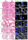
Virus detection in the respiratory system by FISH. HE stained lung tissue and representative image sections showing FISH co-visualization of RNA sequences either of SARS-CoV-2 S gene genomic RNA (green, arrowhead) and ACE2 (red) or SARS-CoV-2 antisense strand RNA as an indicator of replicating virus (green, arrowhead) and TMPRSS2 (red). Morphological details are shown in regions of the alveolus (A, alveolar pneumocytes), endothelium in the alveolar wall (B), endothelial cells adjacent to an embolus (C), hyaline membrane (D), squamous metaplasia (E, metaplastic epithelium), and trachea (F, respiratory epithelium). Scale bars represent 200, 50 and 10 µm, respectively.
Figure 5
Figure 5
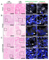
Virus detection in heart and lymphoid organ autopsy tissue by FISH. HE stained tissue and representative image sections showing FISH co-visualization of RNA sequences either of SARS-CoV-2 S gene genomic RNA (green, arrowhead) and ACE2 (red) or SARS-CoV-2 antisense strand RNA indicating replicating virus (green, arrowhead) and TMPRSS2 (red) in the heart (A, cardiomyocyte), tonsil (B, upper panel: capillary with endothelial lining, surrounded by lymphocytes/immune cells; lower panel: local small salivary gland epithelium) and lymph node (C, upper panel: capillary with endothelial lining; lower panel: lymphocytes/immune cells). Scale bars represent 200, 50 and 20 µm, respectively.
Figure 6
Figure 6
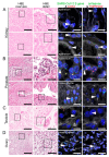
Virus detection in urogenital tract autopsy tissue by FISH. HE stained tissue and representative image sections showing FISH co-visualization of RNA sequences either of SARS-CoV-2 S gene genomic RNA (green, arrowhead) and ACE2 (red) or SARS-CoV-2 antisense strand RNA as an indicator of replicating virus (green, arrowhead) and TMPRSS2 (red) in the kidney (A, upper panel: glomerular visceral epithelial cells/podocytes; lower panel: tubular epithelial cells), prostate (B, upper panel: glandular epithelial cells; lower panel: vascular smooth muscle cells), testicle (C, germinal epithelium) and ovary (D, mesenchymal stromal cells). Scale bars represent 200, 50 and 20 µm (A,C,D) or 10 µm (B), respectively.
Figure 7
Figure 7
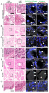
Virus detection in endocrine and gastrointestinal organs and skeletal muscle autopsy tissue by FISH. HE stained tissue and representative image sections showing FISH co-visualization of RNA sequences either of SARS-CoV-2 S gene genomic RNA (green, arrowhead) and ACE2 (red) or SARS-CoV-2 antisense strand RNA indicating replicating virus (green, arrowhead) and TMPRSS2 (red) in the thyroid (A, upper panel: follicular epithelium; lower panel: vascular endothelium), adrenal gland (B, glandular epithelium), salivary gland (C, acini), small bowel (D, crypt epithelium), pancreas (E, acinar epithelium), skeletal muscle (F, skeletal muscle cell) and skin (G). Scale bars represent 200, 50 and 10 µm (A,C,F) or 20 µm (B,D,E), respectively.
Figure 8
Figure 8
Immunohistochemical staining of SARS spike glycoprotein. IHC staining of SARS spike glycoprotein (#Ab272420) in the lung autopsies collected from patients without any respiratory disease (A, Healthy; insert: respiratory epithelial cells) and patients infected with COVID-19 (B, COVID-19; insert: respiratory epithelial cells), influenza (C, Influenza; insert: respiratory epithelial cells) and non-infectious diffuse alveolar damage (D, Non-infect.DAD; insert: cell with features of an alveolar macrophage) with apparently (false-) positive staining (arrowhead). Scale bars represent 40 and 10 µm (insert).

References

    1. Cabrera Martimbianco A.L., Pacheco R.L., Bagattini A.M., Riera R. Frequency, signs and symptoms, and criteria adopted for long COVID: A systematic review. Int. J. Clin. Pract. 2021:e14357. doi: 10.1111/ijcp.14357. - DOI - PMC - PubMed
    1. Wei J.F., Huang F.Y., Xiong T.Y., Liu Q., Chen H., Wang H., Huang H., Luo Y.C., Zhou X., Liu Z.Y., et al. Acute myocardial injury is common in patients with COVID-19 and impairs their prognosis. Heart. 2020;106:1154–1159. doi: 10.1136/heartjnl-2020-317007. - DOI - PMC - PubMed
    1. Sardu C., Gambardella J., Morelli M.B., Wang X., Marfella R., Santulli G. Hypertension, Thrombosis, Kidney Failure, and Diabetes: Is COVID-19 an Endothelial Disease? A Comprehensive Evaluation of Clinical and Basic Evidence. J. Clin. Med. 2020;9:1417. doi: 10.3390/jcm9051417. - DOI - PMC - PubMed
    1. Matarese A., Gambardella J., Sardu C., Santulli G. miR-98 Regulates TMPRSS2 Expression in Human Endothelial Cells: Key Implications for COVID-19. Biomedicines. 2020;8:462. doi: 10.3390/biomedicines8110462. - DOI - PMC - PubMed
    1. V’Kovski P., Kratzel A., Steiner S., Stalder H., Thiel V. Coronavirus biology and replication: Implications for SARS-CoV-2. Nat. Rev. Microbiol. 2021;19:155–170. doi: 10.1038/s41579-020-00468-6. - DOI - PMC - PubMed

Publication types