. 2021 Jul 31;10(8):1946.
doi: 10.3390/cells10081946.

Identification of Novel Cathepsin B Inhibitors with Implications in Alzheimer's Disease: Computational Refining and Biochemical Evaluation

Affiliations

Identification of Novel Cathepsin B Inhibitors with Implications in Alzheimer's Disease: Computational Refining and Biochemical Evaluation

Nitin Chitranshi et al. Cells. .

Abstract

Amyloid precursor protein (APP), upon proteolytic degradation, forms aggregates of amyloid β (Aβ) and plaques in the brain, which are pathological hallmarks of Alzheimer's disease (AD). Cathepsin B is a cysteine protease enzyme that catalyzes the proteolytic degradation of APP in the brain. Thus, cathepsin B inhibition is a crucial therapeutic aspect for the discovery of new anti-Alzheimer's drugs. In this study, we have employed mixed-feature ligand-based virtual screening (LBVS) by integrating pharmacophore mapping, docking, and molecular dynamics to detect small, potent molecules that act as cathepsin B inhibitors. The LBVS model was generated by using hydrophobic (HY), hydrogen bond acceptor (HBA), and hydrogen bond donor (HBD) features, using a dataset of 24 known cathepsin B inhibitors of both natural and synthetic origins. A validated eight-feature pharmacophore hypothesis (Hypo III) was utilized to screen the Maybridge chemical database. The docking score, MM-PBSA, and MM-GBSA methodology was applied to prioritize the lead compounds as virtual screening hits. These compounds share a common amide scaffold, and showed important interactions with Gln23, Cys29, His110, His111, Glu122, His199, and Trp221. The identified inhibitors were further evaluated for cathepsin-B-inhibitory activity. Our study suggests that pyridine, acetamide, and benzohydrazide compounds could be used as a starting point for the development of novel therapeutics.

Keywords: 3D pharmacophore; Alzheimer’s disease; cathepsin B; docking; molecular dynamics; virtual screening.


Conflict of interest statement

The authors declare no conflict of interest.

Figures

Figure 1
Figure 1
Previously reported inhibitors of cathepsin B, with IC50 in µM.
Figure 2
Figure 2
Two-dimensional structures of compound datasets from natural origin (Part 1) and synthetic origin (Part 2). The compound numbers and IC50 values are shown at the bottom of the respective compounds. The training sets are denoted with asterisks.
Figure 2
Figure 2
Two-dimensional structures of compound datasets from natural origin (Part 1) and synthetic origin (Part 2). The compound numbers and IC50 values are shown at the bottom of the respective compounds. The training sets are denoted with asterisks.
Figure 3
Figure 3
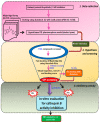
Schematic representation of ligand-based virtual screening of cathepsin B inhibitors. The study was performed in 3 different steps: Step 1, data collection: The dataset of 61 ligands was docked in cathepsin B enzyme (PDBID: 1CSB), and then used to generate a 3D pharmacophore, followed by screening of the Maybridge library. Step 2, hypothesis, and screening: Maybridge hits were subjected to fast docking using AutoDock-Vina, screening using the structural interaction fingerprint (SIFt) approach, and re-docking of the Maybridge virtual screen hits. Step 3, inhibitory activity: Biological evaluation of virtual screening hits against cathepsin B inhibitory activity and molecular dynamics; MM-PBSA and MM-GBSA were used to predict binding free energy components.
Figure 4
Figure 4
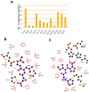
The key residues of the cathepsin B protein involved in interaction studies. (A) Dataset of 61 known cathepsin B inhibitors. LigPlot+ (EMBL-EBI, v2.2, Hinxton, Cambridge, UK) analysis results represent two-dimensional protein–ligand interactions for highly active compounds of (B) natural and (C) synthetic origins. Hydrogen bonds are shown in green dotted lines, while residues interacting by hydrophobic interactions are represented in red.
Figure 5
Figure 5
LigandScout was used to generate a three-dimensional pharmacophore model. (A) An eight-feature hypothesis (Hypo III) and its geometric constraints. Yellow indicates hydrophobic (HY), green indicates hydrogen bond donor (HBD), and red indicates hydrogen bond acceptor (HBA). (B) The best pharmacophore model (Hypo III) aligned to the training set’s most active molecule (compound S1; IC50 0.00224 µM) and (C) the inactive molecule compound N8 (IC50 125 µM). HY: hydrophobic, yellow; HBD: hydrogen bond donor, green; HBA: hydrogen bond acceptor, red.
Figure 6
Figure 6
(A) Cathepsin B and ligand CA030 interaction determined through LigPlot. (B) Virtual screen ranking of the Maybridge compound library. A total of 1728 screened Maybridge compounds were docked to cathepsin B using AutoDock-Vina, then ranked by predicted binding energy. The plot shows the number of active compounds retrieved versus the total number selected (blue line for AutoDock 4.2 and green line for AutoDock-Vina). The grey line indicates the number of active molecules that would be expected to be returned based on a random selection of compounds.
Figure 7
Figure 7
Screening based on structural interaction fingerprints (SIFts). (A) An illustrative representing the SIFt methodology. Fingerprints generated based on binding modes or pairwise interactions (H-bonds and vdW) formed between the proposed docking ligand conformation and a receptor. Step 1: identify the key binding residues of the receptor protein in the complex; step 2: represent each key residue by a bit string (contact, main chain (MC), side chain (SC), polar, non-polar, hydrogen bond acceptor (HBA), and hydrogen bond donor (HBD)) according to the kind of interaction at that residue; step 3: concatenate 7-bit strings of all key residues to form a unique fingerprint, called an SIFt. (B) Dendrogram derived from agglomerative hierarchical clustering of SIFts of known cathepsin B inhibitors and virtual screening hits. Tanimoto similarity coefficient was used to calculate the similarity between the SIFts. (C) The key residues of the cathepsin B protein involved in interaction with virtual screening hit molecules.
Figure 8
Figure 8
Binding modes of lead hits obtained after virtual screening. (A) BTB03075 ligand in red, cathepsin B protein surface view (golden yellow); (a’) Maybridge BTB03075 ligand demonstrating interaction with cathepsin B protein key amino acid residues. (B) KM02922 ligand in yellow, cathepsin B protein surface view (green); (b’) Maybridge KM02922 ligand demonstrating interaction with cathepsin B protein key amino acid residues. (C) RF02795 ligand in blue, cathepsin B protein surface view (purple); (c’) Maybridge RF02795 ligand demonstrating interaction with cathepsin B protein key amino acid residues.
Figure 9
Figure 9
Molecular dynamic simulation performed for 20 ns. (A) The flexibility in the ligand–protein complex was examined by the root-mean-square fluctuation (RMSF) in each receptor–ligand complex. Evolution over time of the root-mean-square deviation (RMSD) of (B) binding site residues, (C) ligands, and (D) proteins.
Figure 10
Figure 10
The activity of virtually screened molecules using a cathepsin B inhibitory screening assay kit. Dose-dependent inhibition of cathepsin B activity by the virtually screened molecules. F-F-FMK was used as a standard inhibitor of cathepsin B activity (positive control, 10 μM concentration), represented as inhibitor control (IC), whereas the solvent with enzyme only (negative controls) is represented as enzyme control (EC). Experiments were performed in triplicate. Data are represented as the mean ± SEM.

References

    1. Bogdanovic N., Hansson O., Zetterberg H., Basun H., Ingelsson M., Lannfelt L., Blennow K. Alzheimer’s disease—The most common cause of dementia. Lakartidningen. 2020;117 - PubMed
    1. Marelli C., Hourregue C., Gutierrez L.A., Paquet C., de Menjot Champfleur N., De Verbizier D., Jacob M., Dubois J., Maleska A.M., Hirtz C., et al. Cerebrospinal Fluid and Plasma Biomarkers do not Differ in the Presenile and Late-Onset Behavioral Variants of Frontotemporal Dementia. J. Alzheimers Dis. 2020;74:903–911. doi: 10.3233/JAD-190378. - DOI - PubMed
    1. Wilson R.S., Segawa E., Boyle P.A., Anagnos S.E., Hizel L.P., Bennett D.A. The natural history of cognitive decline in Alzheimer’s disease. Psychol. Aging. 2012;27:1008–1017. doi: 10.1037/a0029857. - DOI - PMC - PubMed
    1. Gaugler J., James B., Johnson T., Marin A., Weuve J., Assoc A.S. 2019 Alzheimer’s disease facts and figures. Alzheimers Dement. 2019;15:321–387. doi: 10.1016/j.jalz.2019.01.010. - DOI
    1. Gupta V., Gupta V.B., Chitranshi N., Gangoda S., Vander Wall R., Abbasi M., Golzan M., Dheer Y., Shah T., Avolio A., et al. One protein, multiple pathologies: Multifaceted involvement of amyloid beta in neurodegenerative disorders of the brain and retina. Cell Mol. Life Sci. 2016;73:4279–4297. doi: 10.1007/s00018-016-2295-x. - DOI - PMC - PubMed

Publication types

MeSH terms