Review
. 2021 Jul 31;10(8):1957.
doi: 10.3390/cells10081957.

Insights on the Regeneration Potential of Müller Glia in the Mammalian Retina

Affiliations
Review

Insights on the Regeneration Potential of Müller Glia in the Mammalian Retina

Ahmed Salman et al. Cells. .

Abstract

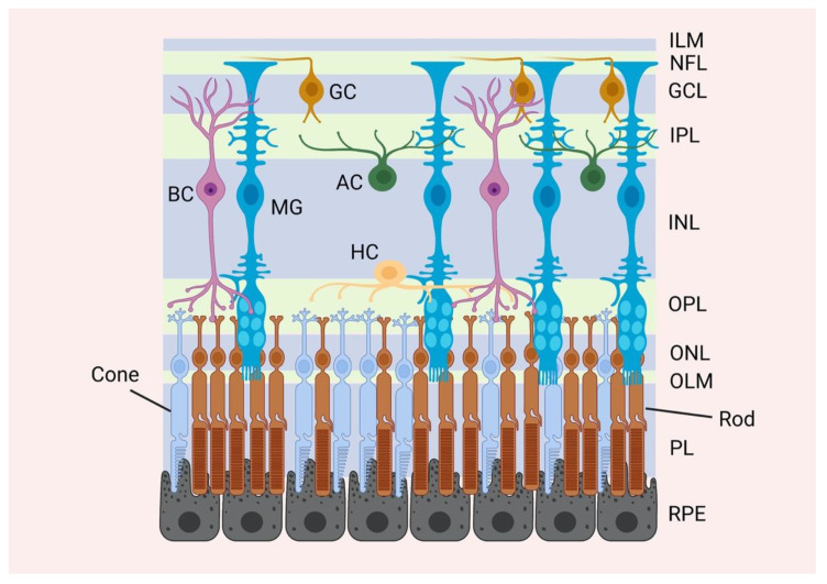
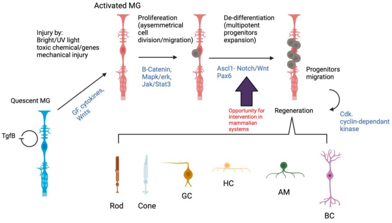
Müller glia, the major glial cell types in the retina, maintain retinal homeostasis and provide structural support to retinal photoreceptors. They also possess regenerative potential that might be used for retinal repair in response to injury or disease. In teleost fish (such as zebrafish), the Müller glia response to injury involves reprogramming events that result in a population of proliferative neural progenitors that can regenerate the injured retina. Recent studies have revealed several important mechanisms for the regenerative capacity of Müller glia in fish, which may shed more light on the mechanisms of Müller glia reprogramming and regeneration in mammals. Mammalian Müller glia can adopt stem cell characteristics, and in response to special conditions, be persuaded to proliferate and regenerate, although their native regeneration potential is limited. In this review, we consider the work to date revealing the regenerative potential of the mammalian Müller glia and discuss whether they are a potential source for cell regeneration therapy in humans.

Keywords: Müller glia; differentiation; proliferation; regeneration potential; reprogramming; retinal regeneration; stem cells.


Conflict of interest statement

The authors declare no conflict of interest.

Figures

Figure 1
Figure 1
The mammalian retinal anatomy. A representative illustration of the major retinal layers and cell types. The mammalian retina is divided into 3 main laminar layers: the outer nuclear layer (ONL), the inner nuclear layer (INL) and the ganglion cell layer (GCL). There are six different retinal neuronal cell types and one glial cell type distributed within the nuclear layers: the rod and cone photoreceptor cell bodies are located in the ONL, whereas the cell bodies of the bipolar (BC), horizontal (HC), amacrine (AC) and Müller (MG) cells are located in the GCL. The cell bodies of the ganglion cells (GC) are located in the GCL. The processes of the different cells are extended into two plexiform layers. Processes from the photoreceptor cells are extended into the outer plexiform layer (OPL) to form synapses with the retinal neurones. Processes from the bipolar, horizontal, amacrine as well as Müller cells are extended into the inner plexiform layer (IPL). The axons of the ganglion cells are directed into the optic nerve through the nerve fibre layer (NFL). Müller glia span the length of the retina from the outer to the inner limiting membranes (OLM and ILM, respectively).
Figure 2
Figure 2
Müller glia regeneration-associated molecular cascades in zebrafish. A hypothetical pathway representing Müller glia activation in response to injury and the sequence of events that leads to retinal regeneration in zebrafish (black text). Müller glia are quiescent in absence of injury, a process regulated by TgfB/Smad pathway. Upon injury, Müller glia are encouraged start reprogramming and enter the cell cycle in a complex of events involving B-catenin, Mapk/erk and Jak/Stat3 signalling pathways, which stimulate proliferation. These pathways stimulate injury-dependant Ascl-1 expression, which regulates the generation of Müller glia-l-derived progenitor cells (MGPCs). Those sequences of events also occur in mammalian systems. However, the MGPCs in zebrafish retina expand and migrate to regenerate the injured retina. This regeneration potential is inefficient in mammalian systems. Intervention at key steps in the zebrafish Müller glia reprogramming might stimulate the regeneration potential in mammalian Müller glia (purple arrow).

References

    1. Enoch J., McDonald L., Jones L., Jones P., Crabb D.P. Evaluating Whether Sight Is the Most Valued Sense. JAMA Ophthalmol. 2019;137:1317–1320. doi: 10.1001/jamaophthalmol.2019.3537. - DOI - PMC - PubMed
    1. Singh M.S., Park S.S., Albini T.A., Canto-Soler M.V., Klassen H., MacLaren R.E., Takahashi M., Nagiel A., Schwartz S.D., Bharti K. Retinal stem cell transplantation: Balancing safety and potential. Prog. Retin. Eye Res. 2020;75:100779. doi: 10.1016/j.preteyeres.2019.100779. - DOI - PMC - PubMed
    1. Trapani I., Auricchio A. Has retinal gene therapy come of age? From bench to bedside and back to bench. Hum. Mol. Genet. 2019;28:R108–R118. doi: 10.1093/hmg/ddz130. - DOI - PMC - PubMed
    1. Kantor A., McClements M.E., MacLaren R.E. CRISPR-Cas9 DNA Base-Editing and Prime-Editing. Int. J. Mol. Sci. 2020;21:6240. doi: 10.3390/ijms21176240. - DOI - PMC - PubMed
    1. McClements M.E., Staurenghi F., MacLaren R.E., Cehajic-Kapetanovic J. Optogenetic Gene Therapy for the Degenerate Retina: Recent Advances. Front. Neurosci. 2020;14:570909. doi: 10.3389/fnins.2020.570909. - DOI - PMC - PubMed

Publication types