Review
. 2021 Aug 10;10(8):2045.
doi: 10.3390/cells10082045.

Diversity of Adult Neural Stem and Progenitor Cells in Physiology and Disease

Affiliations
Review

Diversity of Adult Neural Stem and Progenitor Cells in Physiology and Disease

Zachary Finkel et al. Cells. .

Abstract

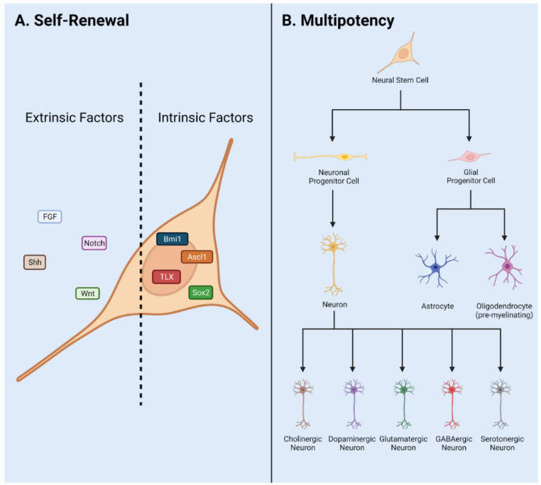
Adult neural stem and progenitor cells (NSPCs) contribute to learning, memory, maintenance of homeostasis, energy metabolism and many other essential processes. They are highly heterogeneous populations that require input from a regionally distinct microenvironment including a mix of neurons, oligodendrocytes, astrocytes, ependymal cells, NG2+ glia, vasculature, cerebrospinal fluid (CSF), and others. The diversity of NSPCs is present in all three major parts of the CNS, i.e., the brain, spinal cord, and retina. Intrinsic and extrinsic signals, e.g., neurotrophic and growth factors, master transcription factors, and mechanical properties of the extracellular matrix (ECM), collectively regulate activities and characteristics of NSPCs: quiescence/survival, proliferation, migration, differentiation, and integration. This review discusses the heterogeneous NSPC populations in the normal physiology and highlights their potentials and roles in injured/diseased states for regenerative medicine.

Keywords: NG2+ cells; central nervous system (CNS); ependymal cells; neural stem and progenitor cells (NSPC); neurodegenerative diseases; regenerative medicine; retina injury; spinal cord injury (SCI); traumatic brain injury (TBI).


Conflict of interest statement

The authors declare no conflict of interest.

Figures

Figure 1
Figure 1
NSPC characteristics in adult mammals. (A) Self renewal requires input via extrinsic and intrinsic factors. These include signaling pathways Notch, Wnt, and Shh, and transcription factors Sox2, Ascl1, Bmi1, Tlx, and neurotransmitters and neurotrophic/trophic growth factors. (B) Multipotency allows NSPCs to differentiate into a variety of cell fates such as Neurons, Astrocytes, and Oligodendrocytes. Adapted from Navarro Quiroz et al., 2018 [6].
Figure 2
Figure 2
NSPC Niche in mammals: the SVZ and SGZ in the brain (A); the ependymal cells and NG2 cells in the spinal cord (B); and the base of the optic nerve, the Müller glia, and the pigment epithelium in the retina (C). AC, anterior chamber; CSF, cerebrospinal fluid; PC, posterior chamber; SVZ, subventricular zone; SGZ, subgranular zone. Adapted from Cutler and Kokovay, 2020 [26] (A); Sabelström et al., 2014 [27], Andreotti et al., 2019; Picoli et al., 2019 [28,29] (B); Yoshida et al., 2000 [30] (C).
Figure 3
Figure 3
Utilities of the Notch1CR2-GFP transgenic mouse line in SCI and TBI models. (A) Notch1CR2-GFP transgenic mouse model labels NSPCs in the CNS. (B) Adult NSPCs in the brain proliferate in the acute phase of TBI and differentiate into neurons in the chronic phase of TBI. (C) In the injured spinal cord, Gsx1 expression promotes adult NSPC proliferation and preferential differentiation into excitatory interneurons and inhibits astrocytes and glial scar formation after injury. Adapted from Tzatzalos, et al., 2012 [81] (A), Anderson et al., 2020 [80] (B) and Patel et al., 2021 [79] (C).
Figure 4
Figure 4
Behavior of ependymal cells and NG2+ cells in animal models of SCI. In normal physiology, the ependymal cells lining the wall of the central canal are largely quiescent, while NG2+ cells are ubiquitously distributed throughout the grey and white matter of the spinal cord. In contusion SCI, the ependymal cell layer is not damaged, but may increase proliferation and differentiation potential. In the hemisection model, the ependymal cell layer is damaged and ependymal cells/NG2+ cells are activated by injury. In stab SCI, the ependymal cell layer is damaged and contributes greatly to glial scar formation. Adapted from Sabelström et al., 2014 [27], Hackett et al., 2016 [103], and Picoli et al., 2019 [29].

References

    1. Kawaguchi A. Neuronal Delamination and Outer Radial Glia Generation in Neocortical Development. Front. Cell Dev. Biol. 2020;8:623573. doi: 10.3389/fcell.2020.623573. - DOI - PMC - PubMed
    1. Molofsky A.V., Pardal R., Iwashita T., Park I.K., Clarke M.F., Morrison S.J. Bmi-1 dependence distinguishes neural stem cell self-renewal from progenitor proliferation. Nature. 2003;425:962–967. doi: 10.1038/nature02060. - DOI - PMC - PubMed
    1. Bond A.M., Berg D.A., Lee S., Garcia-Epelboim A.S., Adusumilli V.S., Ming G.L., Song H. Differential Timing and Coordination of Neurogenesis and Astrogenesis in Developing Mouse Hippocampal Subregions. Brain Sci. 2020;10:909. doi: 10.3390/brainsci10120909. - DOI - PMC - PubMed
    1. Miller F.D., Gauthier A.S. Timing is everything: Making neurons versus glia in the developing cortex. Neuron. 2007;54:357–369. doi: 10.1016/j.neuron.2007.04.019. - DOI - PubMed
    1. Altman J. Are new neurons formed in the brains of adult mammals? Science. 1962;135:1127–1128. doi: 10.1126/science.135.3509.1127. - DOI - PubMed

Publication types