. 2021 Aug 10;10(8):2047.
doi: 10.3390/cells10082047.

Secretory Vesicles Are the Principal Means of SARS-CoV-2 Egress

Affiliations

Secretory Vesicles Are the Principal Means of SARS-CoV-2 Egress

Sébastien Eymieux et al. Cells. .

Abstract

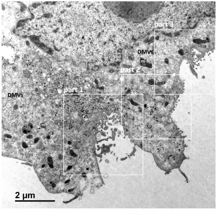
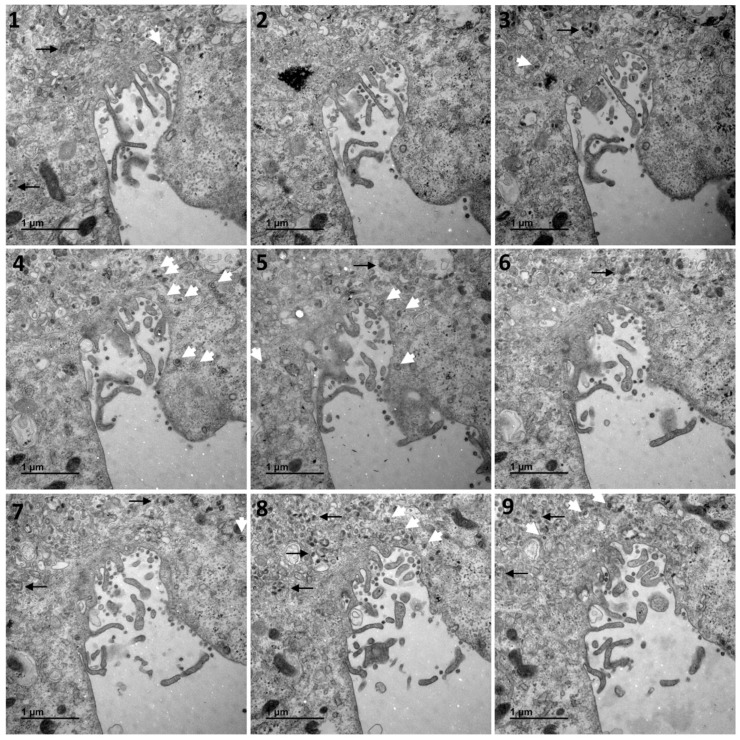
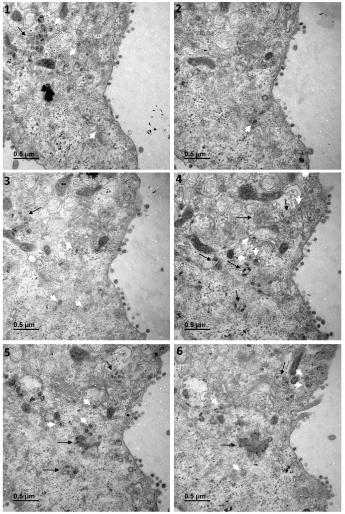
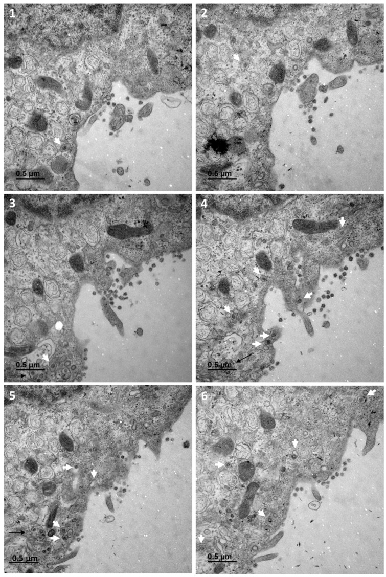
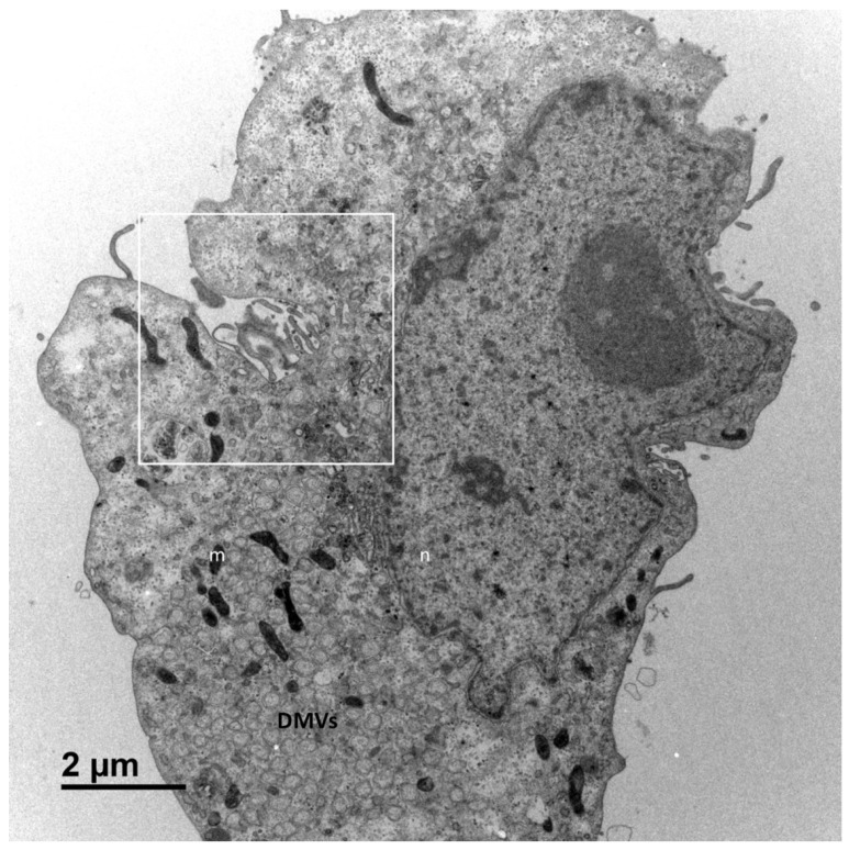
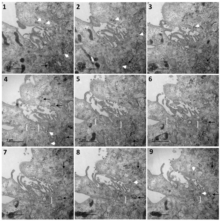
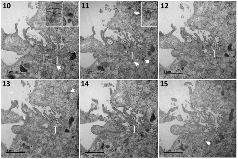
The mechanisms of severe acute respiratory syndrome coronavirus 2 (SARS-CoV-2) egress, similar to those of other coronaviruses, remain poorly understood. The virus buds in intracellular compartments and is therefore thought to be released by the biosynthetic secretory pathway. However, several studies have recently challenged this hypothesis. It has been suggested that coronaviruses, including SARS-CoV-2, use lysosomes for egress. In addition, a focused ion-beam scanning electron microscope (FIB/SEM) study suggested the existence of exit tunnels linking cellular compartments rich in viral particles to the extracellular space resembling those observed for the human immunodeficiency (HIV) in macrophages. Here, we analysed serial sections of Vero cells infected with SARS-CoV-2 by transmission electron microscopy (TEM). We found that SARS-CoV-2 was more likely to exit the cell in small secretory vesicles. Virus trafficking within the cells involves small vesicles, with each generally containing a single virus particle. These vesicles then fuse with the plasma membrane to release the virus into the extracellular space. This work sheds new light on the late stages of the SARS-CoV-2 infectious cycle of potential value for guiding the development of new antiviral strategies.

Keywords: COVID-19; SARS-CoV-2; coronavirus; serial sections; transmission electron microscopy; virus egress; virus release.


Conflict of interest statement

The authors declare no conflict of interest.

Figures

Figure 1
Figure 1
Vero cell at 10 h post-infection with SARS-CoV-2. This cell (cell A) is representative and shows the various virus release situations observed. The three areas delimited by white squares designate the regions of the cell analysed by the examination of serial sections in subsequent Figure 2, Figure 3 and Figure 4: a region characterised by a large cytoplasm invagination rich in plasma membrane microvilli (part 1); a region in which the virus is released and the plasma membrane does not exhibit particular protrusion (part 2); and a region in which the situation is intermediate, with small plasma membrane microvilli at the cell surface (part 3). The cellular area between the cytoplasm invagination and the nucleus (n) contains a large number of viral particles. This area is flanked on both sides by numerous double-membrane vesicles (DMVs) and mitochondria (m).
Figure 2
Figure 2
Serial TEM sections of cell A part 1, a region characterised by a large cytoplasm invagination rich in plasma membrane microvilli. Large intracellular vesicles rich in viral particles (thin black arrows) and small vesicles each con-taining a single viral particle (large white arrows) are shown on each serial section. These electron micrographs of a series of 9 TEM sections (19) are part of a larger series (11 sections) provided at high resolution in the Supplementary Materials. The numbers on the panels indicate the successive serial TEM sections.
Figure 3
Figure 3
Serial TEM sections of cell A part 2, a region in which the virus is released at the plasma membrane without any particular protrusion. Large intracellular vesicles rich in viral particles (thin black arrows) and small vesicles each con-taining a single viral particle (large white arrows) are shown on each serial section. These electron micrographs of a series of 6 TEM sections (16) are part of a larger series (7 sections) provided at high resolution in the Supplementary Materials. The numbers on the panels indicate the successive serial TEM sections.
Figure 4
Figure 4
Serial TEM sections of cell A part 3, a region in which the situation is intermediate to those observed on part 1 and 2, with small plasma membrane microvilli at the cell surface. Large intracellular vesicles rich in viral particles (thin black arrows) and small vesicles each containing a single viral particle (large white arrows) are shown on each serial section. The high-resolution electron micrographs for this series can be consulted in the Supplementary Materials. The numbers on the panels (16) indicate the successive serial TEM sections.
Figure 5
Figure 5
Vero cell at 10 h post-infection with SARS-CoV-2. This cell (cell B) is representative of a frequently observed viral release situation characterised by a large cytoplasm invagination rich in plasma membrane microvilli (area designated by the white square, analysed by the examination of serial TEM sections in Figure 6). The cellular area between the cytoplasm invagination and the nucleus (n) contains a large number of viral particles. This area is close to numerous double-membrane vesicles (DMVs) and mitochondria (m).
Figure 6
Figure 6
Serial TEM sections of the previous area of cell B, characterised by a large cytoplasm invagination rich in plasma membrane microvilli. Large intracellular vesicles rich in viral particles (thin black arrows) and small vesicles each con-taining a single viral particle (large white arrows) are shown on each serial section. The structure identified in white brackets from sections 4 to 12 appears to be an invagination of the plasma membrane, forming a very thin extracellular space between two cellular microvilli. These electron micrographs of a series of 12 TEM sections are part of a larger series (24 sections) provided at high resolution in the Supplementary Materials. Viral particles are released into the thin extra-cellular space between two cellular microvilli through small secretory vesicles each containing a single virus particle (thin white boxes in sections 10 and 11, also shown in insets). On section 10, another viral release event involving a small se-cretory vesicle containing a single viral particle can be visualised (thin white box in the upper part of the image, also shown in the inset). The numbers on the panels (115) indicate the successive serial TEM sections.
Figure 6
Figure 6
Serial TEM sections of the previous area of cell B, characterised by a large cytoplasm invagination rich in plasma membrane microvilli. Large intracellular vesicles rich in viral particles (thin black arrows) and small vesicles each con-taining a single viral particle (large white arrows) are shown on each serial section. The structure identified in white brackets from sections 4 to 12 appears to be an invagination of the plasma membrane, forming a very thin extracellular space between two cellular microvilli. These electron micrographs of a series of 12 TEM sections are part of a larger series (24 sections) provided at high resolution in the Supplementary Materials. Viral particles are released into the thin extra-cellular space between two cellular microvilli through small secretory vesicles each containing a single virus particle (thin white boxes in sections 10 and 11, also shown in insets). On section 10, another viral release event involving a small se-cretory vesicle containing a single viral particle can be visualised (thin white box in the upper part of the image, also shown in the inset). The numbers on the panels (115) indicate the successive serial TEM sections.

References

    1. Boni M.F., Lemey P., Jiang X., Lam T.T., Perry B.W., Castoe T.A., Rambaut A., Robertson D.L. Evolutionary origins of the SARS-CoV-2 sarbecovirus lineage responsible for the COVID-19 pandemic. Nat. Microbiol. 2020;5:1408–1417. doi: 10.1038/s41564-020-0771-4. - DOI - PubMed
    1. World Health Organization WHO Coronavirus Disease (COVID-19) Dashboard. [(accessed on 17 May 2021)]; Available online: https://covid19.who.int/
    1. Al-Horani R.A., Kar S., Aliter K.F. Potential anti-COVID-19 therapeutics that block the early stage of the viral life cycle: Structures, mechanisms, and clinical trials. Int. J. Mol. Sci. 2020;21:5224. doi: 10.3390/ijms21155224. - DOI - PMC - PubMed
    1. Blanchard E., Roingeard P. Virus-induced double-membrane vesicles. Cell. Microbiol. 2015;17:45–50. doi: 10.1111/cmi.12372. - DOI - PMC - PubMed
    1. Snijder E.J., Limpens R.W.A.L., de Wilde A.H., de Jong A.W.M., Zevenhoven-Dobbe J.C., Maier H.J., Faas F.F.G.A., Koster A.J., Bárcena M. A unifying structural and functional model of the coronavirus replication organelle: Tracking down RNA synthesis. PLoS Biol. 2020;18:e3000715. doi: 10.1371/journal.pbio.3000715. - DOI - PMC - PubMed

Publication types